

 PDF
PDF ePub
ePub Citation
 Print
Print


Abstract
Positron emission tomography/computed tomography (PET/CT) is valuable for the diagnosis of malignancies. However, PET/CT is unable to discriminate exactly between inflammation and a neoplasm. We report a case of a 50-year-old man with pulmonary paragonimiasis that was suspicious for lung cancer, as detected by PET/CT. The use of PET/CT revealed multilobulated consolidation on the right lung and patchy consolidation on the left lung, with increased fluorodeoxyglucose (FDG) uptake. In addition, the left paraaortic lymph node (LN) and peripancreatic LN showed enlargement with increased FDG uptake. Lung cancer with multiple lymph node metastases was suspected from the increased standardized uptake values (SUV>4.5) determined by PET/CT. We performed wedge resection via video-assisted thoracic surgery (VATS) and found Paragonimus westermani eggs in the involved tissues.
Figures and Tables
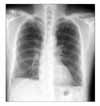
Figure 2
Coronal PET maximum intensity projection image (A) shows multiple increased FDG uptake at whole body. Transverse PET image (upper row), transverse CT lung window image (middle row) and transverse PET/CT image (lower row) show multilobulated consolidation, increased FDG uptake at both upper lobe (B), left upper lobe lingular division (C).
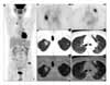
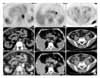
Figure 3
Transverse PET image (upper row), transverse CT image (middle row) and transverse PET/CT image (lower row) show left paraaortic lymph node (A), peripancreatic lymphode enlargement (B) with increased FDG uptake (SUV: 4.64~5.01) and fat infiltration with FDG uptake (SUV: 5.31) at left buttock area (C).
References
1. Lee SH, Chae JI, Hong ST. Synopsis of medical parasitology. 1996. 1st ed. Seoul: Korea Medical Book Publishing Co.
2. Choi JW, Park IS, Shin DH, Park SS, Lee JH. Clinical study of pulmonary paragonimiasis. Tuberc Respir Dis. 1993. 40:274–282.
3. Gould MK, Maclean CC, Kuschner WG, Rydzak CE, Owens DK. Accuracy of positron emission tomography for diagnosis of pulmonary nodules and mass lesions. JAMA. 2001. 285:914–924.
4. Patz EF Jr, Lowe VJ, Hoffman JM, Paine SS, Burrowes P, Coleman RE, et al. Focal pulmonary abnormalities: evaluation with F-18 fluorodeoxyglucose PET scanning. Radiology. 1993. 188:487–490.
5. Patz EF Jr, Goodman PC. Positron emission tomography imaging of the thorax. Radiol Clin North Am. 1994. 32:811–823.
6. Chang JM, Lee HJ, Goo JM, Lee HY, Lee JJ, Chung JK, et al. False positive and false negative FDG-PET scans in various thoracic diseases. Korean J Radiol. 2006. 7:57–69.
7. Yoo IeR, Park HJ, Hyun J, Chung YA, Sohn HS, Chung SK, et al. Two cases of pulmonary paragonimiasis on FDG-PET CT imaging. Ann Nucl Med. 2006. 20:311–315.
8. Watanabe S, Nakamura Y, Kariatsumari K, Nagata T, Sakata R, Zinnouchi S, et al. Pulmonary paragonimiasis mimicking lung cancer on FDG-PET imaging. Anticancer Res. 2003. 23:3437–3440.
9. Goo JM, Im JG, Do KH, Yeo JS, Seo JB, Kim HY, et al. Pulmonary tuberculoma evaluated by means of FDG-PET: findings in 10 cases. Radiology. 2000. 216:117–121.
10. Wilkinson MD, Fulham MJ, McCaughan BC, Constable CJ. Invasive aspergillosis mimicking stage IIIA non-small-cell lung cancer on FDG positron emission tomography. Clin Nucl Med. 2003. 28:234–235.
11. Beggs AD, Hain SF. F-18 FDG-positron emission tomographic scanning and Wegener's granulomatosis. Clin Nucl Med. 2002. 27:705–706.
12. Mukae H, Taniguchi H, Matsumoto N, Iiboshi H, Ashitani J, Matsukura S, et al. Clinicoradiologic features of pleuropulmonary Paragonimus westermani on Kyusyu Island, Japan. Chest. 2001. 120:514–520.
13. DeFrain M, Hooker R. North American paragonimiasis, case report of a severe clinical infection. Chest. 2002. 121:1368–1372.
 XML Download