

 PDF
PDF ePub
ePub Citation
 Print
Print


INTRODUCTION
Myasthenia gravis (MG) is an autoimmune disorder affecting neuromuscular transmission and is characterized by fatigable weakness of voluntary muscles.1 The identification of impaired neuromuscular transmission is an essential step in diagnosing MG.2 Therefore, electrodiagnostic studies (EDx) including repetitive nerve stimulation (RNS) testing and single-fiber electromyography are used to evaluate the electrophysiologic status of the neuromuscular junction.3
Single nerve stimulation may induce several repetitive compound muscle action potentials (R-CMAPs) following the initial CMAP in the presence of certain neuromuscular junction disorders such as organophosphate intoxication, congenital acetylcholinesterase (AChE) deficiency, and slow-channel congenital myasthenic syndrome.4 These R-CMAPs result from repetitive discharge of muscle fibers after a single stimulation and represent the electrophysiologic status of cholinergic neuromuscular hyperactivity.45
R-CMAPs may be induced by AChE inhibitor (AChEI) overdose in patients with MG, and they are rarely seen in MG patients taking a standard dose of AChEI.56 R-CMAPs were found to develop more frequently in MG patients with nicotinic side effects of AChEI than in patients without these side effects.7 In addition, R-CMAPs were frequently observed in muscle-specific tyrosine kinase (MuSK)-antibody-positive MG patients during diagnostic neostigmine testing (NT), and a substantial proportion of the MuSK-antibody-positive MG patients with R-CMAPs could not tolerate AChEI at all due to the cholinergic side effects.5 These findings suggest that the presence of R-CMAPs may predict the side effects and intolerance to oral AChEI by representing the cholinergic activity status in the neuromuscular junction.
In this study we investigated the clinical characteristics of MG patients with R-CMAPs to identify their clinical usefulness in therapeutic decision-making.
METHODS
We retrospectively reviewed the clinical records and electrodiagnostic findings of MG patients who underwent EDx and diagnostic NT from 2007 to 2015 at Yonsei University Hospital. The diagnosis of MG was based on the symptoms and signs of muscle fatigue, decreased responses to low-frequency RNS, high serum levels of anti-acetylcholine-receptor (anti-AChR) antibodies and anti-MuSK antibodies, and the improvement of muscle fatigue after an intramuscular injection of neostigmine. Antibody analyses were performed using commercially available assays (anti-AChR antibodies: Seoul Clinical Laboratories, Seoul, Korea; anti-MuSK antibodies: Athena Diagnostics, Worcester, MA, USA). The test for anti-AChR-binding antibody was considered positive if the value was >0.2 nmol/L. The results of anti-MuSK antibody tests were categorized into negative (<10 titer units), borderline (≥10 and <20 titer units), and positive (≥20 titer units). When patients with MG are evaluated in our electrodiagnostic laboratory, they usually undergo clinical evaluation, RNS testing, and NT. RNS was applied according to previously described methods to the abductor digiti minimi (ADM) and flexor carpi ulnaris (FCU) muscles with ulnar nerve stimulation at the elbow, the orbicularis oculi (OO) and nasalis muscles with facial nerve stimulation, and the trapezius muscle with spinal accessory nerve stimulation using the Neuroscreen system (Toennies, Hoechberg, Germany) or the Schwarzer topas EMG system (Natus, Munich, Germany).8 A decrease in CMAP amplitude of ≥10% was considered to be abnormal. In patients taking pyridostigmine bromide (PB), the EDx was performed at least 12 hours after the last dose. NT was performed after a baseline clinical examination and EDx. The response was evaluated approximately 30 minutes after an intramuscular injection of 0.02 mg/kg neostigmine methylsulfate. The side effects of neostigmine were recorded. At the examiner's discretion, ulnar nerve stimulation recordings on the ADM could be repeated 30–45 minutes after neostigmine injection to confirm the objective electrophysiologic responsiveness to neostigmine methylsulfate.
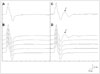
A total of 79 patients with MG underwent RNS testing before NT and ulnar nerve stimulation after NT. We excluded patients who had incomplete medical records or less than 12 months of follow-up after EDx. The patients were classified into two groups according to the development of R-CMAPs on ulnar nerve stimulation after NT. R-CMAPs were defined as a CMAP followed by repetitive discharges that did not exist before NT (Fig. 1). Two neurologists (L.H.S. and P.B.S.) reviewed the results of EDx while blinded to clinical information about the patients, and consensually determined the presence of R-CMAPs in the ADM muscle at normal gain (5 mV/division).
The background information of the patients was obtained from their medical records, including the age at MG symptom onset, sex, MG Foundation of America (MGFA) clinical classification at the first visit, the worst MGFA clinical classification during follow-up, results of anti-AChR and anti-MuSK antibody tests, presence of thymoma, follow-up duration, MGFA postintervention status, highest daily dose of PB, intolerance to PB, and side effects of PB. Intolerance to PB was defined as an inability to take PB due to cholinergic side effects such as severe muscle fasciculation, abdominal cramps, hypersalivation, and blurred vision. The following information was obtained from EDx reports: age at EDx, disease duration, results of RNS testing, MG activities of daily living (MG-ADL) score, baseline quantitative MG score, result of NT, and side effects of neostigmine.
This study was approved by the Institutional Review Board of Severance Hospital, Yonsei University Health System, which waived the requirement for obtaining informed consent from the study subjects due to them remaining anonymous (IRB No. 4-2016-0060).
Statistical analysis
Continuous variables were compared between two groups using the Mann-Whitney U test. The Pearson's chi-square test and Fisher's exact test were used for comparing categorical variables between groups. All statistical analyses were performed using the IBM SPSS Statistics software for Windows version 21.0 (IBM Corporation, Armonk, NY, USA). A two-tailed probability value of p<0.05 was considered statistically significant.
RESULTS
Among the 79 patients who underwent EDx before NT and ulnar nerve stimulation after NT, 71 patients were included in this study. The eight excluded patients comprised seven with less than 12 months of follow-up and one with incomplete medical records. None of these patients exhibited R-CMAPs in baseline RNS testing. After neostigmine injection, R-CMAPs developed in the ADM in 24 of the 71 MG patients.
Clinical manifestations
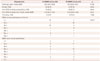
The clinical characteristics of the patients are summarized in Table 1. The age at disease onset and presence of thymoma did not differ significantly between the patients with and without R-CMAPs. The frequency of ocular myasthenia (41.7% vs. 14.9%, p=0.012) and the proportion of females (91.7% vs. 57.4%, p=0.003) were significantly higher in the patients with R-CMAPs than in those without R-CMAPs. The rate of anti-AChR-antibody seropositivity was significantly lower in the patients with R-CMAPs than in those without R-CMAPs (58.3% vs. 85.1%, p=0.012). The anti-MuSK-antibody assay was performed in 17 of the 71 patients, and seropositivity was found in 10 patients, with R-CMAPs developing in 9 of these 10.
Repetitive nerve stimulation and neostigmine testing
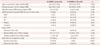
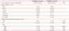
There were no significant differences in the age at the examination, MG-ADL score at the RNS testing, and disease duration between the patients with and without R-CMAPs. The frequency of decreased responses in the baseline RNS testing was significantly lower in the patients with R-CMAPs than in those without R-CMAPs (ADM: 0.0% vs. 61.7%, p<0.001; FCU: 16.7% vs. 72.3%, p<0.001; OO: 50.0% vs. 74.5%, p=0.039; nasalis: 50.0% vs. 87.2%, p=0.001; trapezius: 33.3% vs. 68.1%, p=0.005) (Table 2).
The rate of positive results in the NT was significantly lower in the patients with R-CMAPs than in those without R-CMAPs. Side effects of neostigmine were present in all of the patients with R-CMAPs and in 33 patients without R-CMAPs (100% vs. 70.2%, p=0.002). Twenty-three (95.8%) of the patients with R-CMAPs exhibited muscarinic side effects of neostigmine (abdominal pain in 23 patients and diarrhea in 1). Thirty-two (68.1%) of the patients without R-CMAPs also exhibited muscarinic side effects (abdominal pain in 29, diarrhea in 5, increased salivation in 3, and nausea in 1). Nicotinic side effects of neostigmine were significantly more frequent in the patients with R-CMAPs than in those without R-CMAPs (75.0% vs. 6.4%, p<0.001). Among the 18 patients with R-CMAPs exhibiting nicotinic side effects, 16 had muscle fasciculation, 1 had a squeezing sensation in the neck, and 1 had both side effects. Only three (6.4%) of the patients without R-CMAPs exhibited nicotinic side effects.
Side effects of pyridostigmine bromide
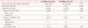
Table 3 lists the side effects of PB. The highest daily dose of PB was lower in the patients with R-CMAPs (240 mg/day vs. 480 mg/day, p<0.001). The frequency of PB intolerance was higher in the patients with R-CMAPs than in those without R-CMAPs (37.5% vs. 0%, p<0.001). The reasons for intolerance were abdominal pain (n=6) and muscle fasciculation (n=3). Side effects of PB were significantly more frequent in the patients with R-CMAPs than in those without R-CMAPs (45.8% vs. 12.8%, p=0.002). Nicotinic side effects of PB were significantly more frequent in the patients with R-CMAPs than in those without R-CMAPs (20.8% vs. 4.3%, p=0.040), whereas the frequency of muscarinic side effects did not differ significantly between the patients with and without R-CMAPs.
The treatment and postintervention status of myasthenia gravis patients
The treatment and postintervention status of the patients are summarized in Table 4. The follow-up duration did not differ significantly between patients with and without R-CMAPs (57.5 months vs. 56.0 months, p=0.316). The frequency of a good treatment response [i.e., postintervention status of complete and stable remission (CSR), pharmacologic remission (PR), or minimal manifestation (MM); MM1, The patient continues to receive some form of immunosuppression but no cholinesterase inhibitors or other symptomatic therapy; MM2, The patient has received only low-dose cholinesterase inhibitors (<120 mg pyridostigmine/day) for at least 1 year; MM3, The patient has received cholinesterase inhibitors or other symptomatic therapy and some form of immunosuppression during the past year) also did not differ between the patients with and without R-CMAPs (CSR: 0.0% vs. 10.6%, p=0.159; PR: 41.7% vs. 27.7%, p=0.920; MM1: 33.3% vs. 12.8%, p=0.058; MM2: 12.5% vs. 14.9%, p=1.000; MM3: 12.5% vs. 31.9%, p=0.075).
DISCUSSION
AChEIs facilitate neuromuscular transmission by inhibiting acetylcholine breakdown at the neuromuscular junction.9 These drugs are used as the first-line treatment of MG and provide temporary relief of symptoms.91011 Although AChEIs are usually tolerated well at standard doses (e.g., up to 60 mg of PB five times per day10), a substantial proportion of MG patients receiving regular treatment with AChEI suffer from its side effects, which can decrease their quality of life.9 Furthermore, a small proportion of MG patients show cholinergic hypersensitivity and cannot tolerate even a low dose of AChEIs.5 In our study, similar to the previous studies, 9 of 71 MG patients did not tolerate oral PB at all, and about one-quarter of 62 MG patients receiving regular treatment with oral PB suffered adverse side effects of PB. In addition, intolerance to PB occurred only in the MG patients with R-CMAPs. The side effects of PB developed more frequently in the MG patients with R-CMAPs than in those without R-CMAPs.
While the intolerance to and the side effects of AChEIs were more frequent in MG patients with R-CMAPs than in those without R-CMAPs, the MGFA postintervention status did not differ significantly between MG patients with and without R-CMAPs, and the response of MG treatment to immunotherapy was good in both groups in the present study. Sometimes AChEIs are poorly tolerated or can lead to clinical worsening in MG patients, especially those who are seropositive for MuSK antibodies,591012 and a cholinergic crisis can occur in severe cases.13 Nine of the MG patients in our study did not tolerate oral PB. Among them, six patients suffered from severe and intolerable side effects during a substantial period due to the inexperience of primary doctors. The side effects disappeared within a few days after discontinuing oral PB in all six patients. They were treated with prednisolone and/or other immunosuppressants and all showed good treatment responses. These observations indicate that therapeutic management with AChEIs should be individualized.
R-CMAPs (i.e., two or more consecutive CMAPs) may be evoked by a single nerve stimulation in the presence of several neuromuscular junction disorders such as organophosphate intoxication and congenital AChE deficiency, and slow-channel congenital myasthenic syndrome.4 In these conditions, the endplate potential (EPP) is prolonged and the amplitude of EPP remains above the muscle fiber action potential threshold for longer than its absolute refractory period.141516 This abnormally prolonged EPP causes re-excitation of the muscle fibers and the generation of R-CMAPs.4 Therefore, R-CMAPs reflect the electrophysiologic status of cholinergic neuromuscular hyperactivity in the neuromuscular junction.5 A previous study of the side effects of AChEI in patients with MG found that patients who experienced nicotinic side effects such as muscle twitching and cramps developed R-CMAPs more frequently than did those without such side effects.7 Another recent study showed that nicotinic side effects of AChEI and R-CMAPs were more frequent in MuSK-antibody-positive than MuSK-antibody-negative MG patients.5 The findings of the previous and present studies suggest that R-CMAPs are associated with the side effects of AChEI and therefore may be used as an electrophysiologic parameter to aid therapeutic decision-making in patients with MG. In addition, checking the presence of R-CMAPs after NT is very easy when performing EDx and NT, which are essential tests for diagnosing MG.
Considering the mechanisms underlying the effects of AChEIs, measuring AChE activity must be a reasonable method for assessing the side effects of AChEIs. However, there is no method available for directly measuring the activity of AChE in effector tissues including skeletal muscles, smooth muscles, and autonomic glands; instead, only the erythrocyte AChE activity can be measured in practice.17 Previous studies of AChEIs found that the level of cholinesterase inhibition—calculated using erythrocyte AChE activity—was not correlated with the extent or severity of its side effects.1118 Therefore, erythrocyte AChE activity is probably not a reliable indicator of AChEI side effects. Polymorphism in the AChE promoter, which has been reported in patients exhibiting hypersensitivity to AChEI, may induce severe and intolerable side effects of AChEI even at low doses.19
Some issues and limitations of the present study must be considered. This was a retrospective study and is unlikely to have been free from selection bias. The frequency of ocular myasthenia and the proportion of females were significantly higher and the frequency of anti-AChR-antibody seropositivity was significantly lower in the patients with R-CMAPs than in those without R-CMAPs. These differences might be influenced by selection bias. However, R-CMAPs develop frequently in MuSK-antibody-positive MG patients, with a marked female predominance.512 Bulbar symptoms appeared frequently in MG patients with R-CMAPs in the present study. Twelve of the 14 MG patients with R-CMAPs were classified as MGFA class IIb, IIIb, or IVb. Although the MuSK-antibody test was not performed in all subjects, it is possible that R-CMAPs were more common in MuSK-antibody-positive MG patients. Therefore, a female predominance and anti-AChR-antibody seronegativity may be characteristic features of MG patients with R-CMAPs. Ocular myasthenia was more frequent in the patients with R-CMAPs. According to our experience, R-CMAPs do not develop in MG patients with a significant neuromuscular transmission failure. In the present study, R-CMAPs occurred only when the CMAP decreased by less than 10% in the ADM. In addition, mild weakness was more common in the patients with R-CMAPs than in those without R-CMAPs (MGFA class II: 78.6% vs. 55.0%, MGFA classes III–V: 21.4% vs. 45.0%, p<0.001). These observations suggest that the high frequency of ocular myasthenia is associated with relatively intact neuromuscular transmission in the ADM in R-CMAP patients. We are planning a future study with a prospective design for predicting the side effects of oral PB.
In conclusion, the side effects of AChEIs and intolerance to oral PB are more common in MG patients with R-CMAPs than in those without R-CMAPs. Furthermore, both MG patients with and without R-CMAPs respond well to immunotherapy. AChEIs should therefore be used carefully in MG patients with R-CMAPs. R-CMAPs after NT may indicate cholinergic neuromuscular hyperactivity to AChEIs and be useful for predicting the intolerance to and side effects of oral PB.
 XML Download