

 PDF
PDF ePub
ePub Citation
 Print
Print


INTRODUCTION
Tuberculosis is caused by the bacteria Mycobacterium tuberculosis and is a global concern worldwide, especially in developing countries.1 Despite the rapid economic development of the Republic of Korea, the prevalence of tuberculosis is high, with an incidence of 108 cases per 100,000 people.12 Tuberculosis meningitis is known to be the most severe form of tuberculosis and is detected in less than 1% of all tuberculosis cases.3 It primarily affects the meninges of the brain and spinal cord along with the adjacent brain parenchyma.1 Tuberculosis meningitis is a devastating disease because half of the affected patients either die or suffer from permanent sequelae.3 Early diagnosis and treatment is important for better prognoses. However, the diagnosis is often problematic despite significant advances in diagnostic techniques, resulting in tuberculosis meningitis often only being diagnosed after a substantial delay.4 There are several reasons for a delayed diagnosis of tuberculosis meningitis. First, the initial presentation of tuberculosis meningitis may be similar to other types of meningitis, and it is especially difficult to differentiate from partially treated bacterial meningitis. Second, smear tests for acid-fast bacillus are positive in only 5–30% of patients with tuberculosis meningitis. In addition, cultures of Mycobacterium tuberculosis from the cerebrospinal fluid (CSF) are positive in approximately 50% of cases, and a result takes several weeks to obtain. The detection of Mycobacterium tuberculosis DNA in the CSF using the polymerase chain reaction (PCR) is a widely used diagnostic method, but its sensitivity is only 56%.1 Thus, simple and rapid predictors for diagnosing tuberculosis meningitis are needed.
Procalcitonin is a peptide consisting of 116 amino acids and is a calcitonin precursor.5 It is usually synthesized in the thyroid gland.6 However, there is evidence that cells of the monocyte-macrophage system are capable of synthesizing procalcitonin, as well as other nonthyroidal tissues, mostly parenchymal cells, when stimulated by bacterial products.5 During a bacterial infection, the production of procalcitonin is induced by tumor necrosis factor alpha (TNF-α) and interleukin 2 (IL-2).7 This makes serum procalcitonin a sensitive marker of severe bacterial infection, and many studies have found serum procalcitonin to be the best marker for differentiating bacterial meningitis from viral meningitis.89 In addition, there is evidence that measuring the level of procalcitonin in the blood is useful for differentiating bacterial pneumonia from tuberculosis and pyogenic spondylodiscitis from tuberculosis spondylodiscitis.27101112 Moreover, the levels of procalcitonin in the blood and pleural fluid differ significantly between patients with tuberculosis and nontuberculosis pleurisy.13 However, no study has investigated whether serum procalcitonin is a useful marker for discriminating between tuberculosis meningitis and bacterial meningitis. Additionally, no published studies have addressed the value of serum procalcitonin as a predictive factor for the prognosis of tuberculosis meningitis.
The aim of this study was to elucidate the potential role of serum procalcitonin in differentiating tuberculosis meningitis from bacterial and viral meningitis, and in predicting the prognosis of tuberculosis meningitis.
METHODS
Participants
This study was approved by the Institutional Review Board at our institution. It had a retrospective study, and involved patients with a clinical suspicion of tuberculosis meningitis admitted to the neurology departments of two tertiary hospitals in Busan, Korea from January 2009 to July 2015. All of the patients had typical clinical histories and laboratory findings of tuberculosis meningitis. In addition, all patients who were admitted to centers with a clinical suspicion of bacterial and viral meningitis during the same period were included as the disease control group for comparisons.
We enrolled patients with tuberculosis meningitis according to the current consensus of the diagnosis and a scoring system, and classified them into definite and probable tuberculosis meningitis.14 The scoring system comprised four parts—clinical criteria, CSF criteria, cerebral imaging criteria, and evidence of tuberculosis elsewhere—and points were given for variables that were present. Patients were judged to have definite tuberculosis meningitis if Mycobacterium tuberculosis was identified in their CSF. Patients had probable tuberculosis meningitis if they scored at least 10 with cerebral imaging results and 12 points without cerebral imaging results.14 In addition, we defined bacterial meningitis based on a combination of the clinical history and laboratory findings. These included 1) clinical features, such as the acute onset of headache, fever, and signs of meningeal irritation; 2) positive CSF findings, including pleocytosis (≥5/mm3, mainly neutrophilic), elevated protein concentration (≥45 mg/dL), a reduced ratio of CSF glucose to serum glucose (≤0.60); 3) a negative CSF stain, culture, or PCR for viruses, mycobacteria, and fungi; and 4) a positive CSF culture, smear, or PCR for bacterial pathogens or a good specific response to antibacterial therapy.15 Moreover, we included patients with viral meningitis. These patients had 1) clinical features, such as the acute onset of headache, fever, and signs of meningeal irritation; 2) no signs of cortical involvement such as altered consciousness, aphasia, or seizures; 3) positive CSF findings including pleocytosis (≥5/mm3, mainly lymphocytic); 4) a negative CSF stain, culture, or PCR for bacteria, mycobacteria, and fungi; and 5) a positive PCR for viral pathogens or full recovery without any specific treatments including antibacterial or antituberculosis therapy.15 We excluded patients whose blood procalcitonin levels had not been measured.
A total of 26 patients with tuberculosis meningitis met the inclusion criteria for this study (Fig. 1). In addition, we included 70 patients with bacterial meningitis and 49 patients with viral meningitis as disease control groups. Only 12 of the 26 patients with tuberculosis meningitis had a positive CSF culture, smear, or PCR for Mycobacterium tuberculosis, and they were classified as definite tuberculosis meningitis. The remaining 14 patients were classified as probable tuberculosis meningitis.14 Of the 70 patients with bacterial meningitis, 41 had a positive CSF culture, smear, or PCR for bacterial pathogens, comprising 22 with Streptococcus species, 7 with Staphylococcus species, 4 with Klebsiella species, 4 with Listeria species, and 4 with other species. Of the 49 patients with viral meningitis, 23 with viral pathogens were identified using PCR of the CSF, comprising 15 with enterovirus, 5 with herpes simplex virus, 2 with herpes zoster virus, and 1 with Epstein-Barr virus.
Measurements
Differences in the demographic and laboratory data among patients with tuberculosis, bacterial, and viral meningitis were analyzed using the demographic profile including age, sex, and diabetes mellitus; blood profile including the white blood cell (WBC) count, platelet count, C-reactive protein (CRP), and procalcitonin; CSF profile including the WBC count, lymphocyte percentage of the total CSF WBC count (lymphocyte percentage), protein, adenosine deaminase (ADA), and the glucose ratio (CSF/blood); and clinical profile including systolic and diastolic blood pressures, heart rate, and score on the Glasgow Coma Scale (GCS) at the times of admission and discharge. We analyzed the blood pressure, heart rate, and blood and CSF profiles that were obtained on the day of admission. In addition, we analyzed the predictive factors for the prognosis of tuberculosis meningitis, which was based on the GCS score at discharge, which was used to divide the patients with tuberculosis meningitis into two groups: good prognosis (GCS score >8) and poor prognosis (GCS score ≤8). Moreover, we analyzed the correlations between the GCS score at discharge and demographic profile including age; blood profile including the WBC count, platelet count, CRP, and procalcitonin; CSF profile including the WBC count, lymphocyte percentage, protein, ADA, and glucose ratio (CSF/blood); and clinical profile including systolic and diastolic blood pressures, heart rate, and GCS score at admission.
Procalcitonin concentrations were measured using an electrical chemiluminescence assay (cobas e 411, Roche Diagnostics, Indianapolis, IN, USA), for which the measurement range was 0.05–200 ng/mL.
Statistical analysis
Comparisons were analyzed using the chi-square test for categorical variables and Student's t-test, Mann-Whitney U-test, analysis of variance, or Kruskal-Wallis test for numerical variables. Categorical variables are presented as frequency and percentage values. Numerical variables conforming to a normal distribution are presented as mean±standard deviation values, while other numerical variables are presented as median and interquartile-range values. In addition, separate bivariate logistic regression models for each tool were used to determine the odds ratios for differentiating tuberculosis meningitis from bacterial meningitis and tuberculosis meningitis with a poor prognosis from that with a good prognosis. To perform multivariate analyses and evaluate the sensitivity and specificity for distinguishing tuberculosis meningitis from bacterial meningitis, we analyzed the clear cutoff values using the receiver operating characteristic (ROC) curve. The data obtained using ROC curves are presented as 95% confidence interval (CI) and standard error (SE) values. Continuous variables were converted into categorical variables by dichotomizing them in accordance with the clear cutoff values in the logistic regression analysis. The correlation analysis was performed using Spearman's rank correlation test. Probability values of p<0.05 were considered to be indicative of statistical significance. We set p<0.017 (0.05/3) as significant when comparing the demographic and laboratory differences among the patients with tuberculosis, bacterial, and viral meningitis with multiple corrections in post-hoc analysis. All statistical tests were performed using MedCalc® (version 13, MedCalc Software, Ostend, Belgium).
RESULTS
Differences in measurements among the total participants
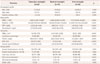
Table 1 compares the demographic and laboratory profiles among the patients with tuberculosis, bacterial, and viral meningitis. The demographic profile including age; the blood profile including the WBC count, platelet count, CRP, and procalcitonin; the CSF profile including the WBC count, lymphocyte percentage, protein, and ADA; and the clinical profile including GCS scores at admission and discharge differed significantly among the patients with tuberculosis, bacterial, and viral meningitis.
In the post-hoc analysis, the patients with tuberculosis meningitis had a higher platelet count in blood and GCS score at admission, and a lower WBC count, CRP, and procalcitonin in the blood, and a lower WBC count, lymphocyte percentage, and ADA in the CSF than those with bacterial meningitis. In addition, the patients with tuberculosis meningitis were older and had higher levels of protein and ADA in the CSF, and lower lymphocyte percentages in the CSF and lower GCS scores at admission and discharge than those with viral meningitis. However, the level of procalcitonin did not differ significantly between patients with tuberculosis meningitis and those with viral meningitis.
Differences in measurements between patients with tuberculosis meningitis and bacterial meningitis
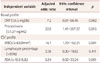
The area under the ROC curve (AUC) was 0.784 (95% CI=0.688–0.862, SE=0.152) for the CRP in the blood, 0.835 (95% CI=0.746–0.903, SE=0.040) for the procalcitonin level in the blood, 0.782 (95% CI=0.686–0.860, SE=0.047) for the WBC count in the CSF, 0.676 (95% CI=0.567–0.772, SE=0.064) for the lymphocyte percentage in the CSF, and 0.767 (95% CI=0.659–0.854, SE=0.061) for the ADA in the CSF. Multiple logistic regression analysis showed that a low level of procalcitonin (≤1.27 ng/mL) in the blood and a low WBC count (<630/mm3) in the CSF were independently significant variables for predicting tuberculosis meningitis (Table 2). The risk of having tuberculosis meningitis with a low serum level of procalcitonin (≤1.27 ng/mL) was at least 22 times higher than the risk of having bacterial meningitis. These variables have very high sensitivity and negative predictive values, but low specificity and positive predictive values. Compared to the WBC count in the CSF, the level of procalcitonin in the blood had a higher sensitivity [96.2% (95% CI=80.4–99.9%) vs. 92.3% (95% CI=74.9–99.1%)], specificity [62.9% (95% CI=50.5–74.1%) vs. 57.1% (95% CI=44.8–68.9%)], positive predictive value [49.0% (95% CI=34.8–63.4%) vs. 44.4% (95% CI=30.9–58.6%)], negative predictive value [97.8% (95% CI=88.2–99.9%) vs. 95.2% (95% CI=83.8–99.4%)], positive likelihood ratio [2.6 (95% CI=1.9–3.5) vs. 2.2 (95% CI=1.6–2.9)], and negative likelihood ratio [0.1 (95% CI=0.0–0.4) vs. 0.1 (95% CI=0.0–0.5)].
Differences in measurements between tuberculosis meningitis patients with good and poor prognoses
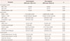
Of the 26 patients with tuberculosis meningitis, 21 had a good prognosis (GCS score >8 at discharge) and 5 had a poor prognosis. The WBC count and procalcitonin in the blood were higher in patients with a poor prognosis than those with a good prognosis (Table 3). The AUC was 0.848 (95% CI=0.653–0.957, SE=0.124) for the WBC count in the blood and 0.876 (95% CI=0.688–0.972, SE=0.097) for the procalcitonin level in the blood. Multiple logistic regression analysis showed that only a high level of procalcitonin (>0.4 ng/mL) in the blood was an independent and significant predictor of a poor prognosis in patients with tuberculosis meningitis (Table 4). The risk of having a poor prognosis in patients with tuberculosis meningitis with a high serum level of procalcitonin (>0.4 ng/mL) was at least 20 times higher than for those with a good prognosis. The level of procalcitonin (r=-0.437, p=0.026), diastolic blood pressure (r=-0.493, p=0.010), and heart rate (r=-0.471, p=0.015) in patients with tuberculosis meningitis were negatively correlated with the GCS score at discharge (Fig. 2A). In addition, there was a positive correlation between the GCS scores at admission and discharge (r=0.439, p=0.025). However, age (r=-0.154, p=0.453), systolic blood pressure (r=-0.267, p=0.187), blood profile including WBC count (r=-0.376, p=0.058), platelet count (r=0.0397, p=0.847), and CRP (r=-0.0515, p=0.803), CSF profile including WBC count (r=0.167, p=0.425), lymphocyte percentage (r=0.029, p=0.897), ADA (r=0.189, p=0.375), and protein (r=0.309, p=0.142), and glucose ratio (CSF/blood) (r=0.154, p=0.474) were not correlated with the GCS score at discharge. In addition, the levels of procalcitonin in patients with bacterial and viral meningitis were not correlated with the GCS score at discharge (r=-0.043, p=0.725 and r=-0.182, p=0.211, respectively) (Fig. 2B and C).
DISCUSSION
This study was the first to investigate the usefulness of the serum procalcitonin level as a diagnostic and prognostic factor for tuberculosis meningitis. We found that the level of procalcitonin in the blood at admission was lower in patients with tuberculosis meningitis than in those with bacterial meningitis. Moreover, we identified that a high level of procalcitonin in the blood at admission was an independent significant predictor of a low GCS score at discharge in patients with tuberculosis meningitis. In addition, the level of procalcitonin in the blood at admission in patients with tuberculosis meningitis was negatively correlated with the GCS score at discharge. These results suggest that serum procalcitonin is a useful marker for the diagnosis and prognosis prediction of tuberculosis meningitis at the initial diagnosis stage.
We found that the blood profile including the WBC count and CRP and the CSF profile including the WBC count, lymphocyte percentage, and ADA differed significantly between patients with tuberculosis and patients with bacterial meningitis. This finding is in agreement with those of previous studies.16 In addition to these findings, we have demonstrated that the level of procalcitonin in the blood differs between patients with tuberculosis meningitis and patients with bacterial meningitis. The high sensitivity and negative predictive value for differentiating the diagnoses of tuberculosis meningitis and bacterial meningitis suggest a supplementary role for serum procalcitonin in the diagnostic exclusion of tuberculosis meningitis from bacterial meningitis in countries where tuberculosis is endemic, such as the Republic of Korea. This result is consistent with previous studies that demonstrated the utility of serum procalcitonin for differentiating pulmonary tuberculosis from bacterial pneumonia.7101112 The reason why the level of procalcitonin in the blood remains relatively low in tuberculosis meningitis is unclear. The cascade of inflammatory cytokines released during a systemic infection may determine the rate and intensity of procalcitonin synthesis and release, thus accounting for differences observed in the blood levels of procalcitonin. This cascade is probably greater in bacterial meningitis than tuberculosis meningitis.12 The elevated levels of TNF-α and interferon-gamma (IFN-γ) produced during mycobacterial infection play key roles in the cellular host response in the immunopathogenesis of tuberculosis meningitis.1 The production of procalcitonin is induced by TNF-α but inhibited by IFN-γ.71718 Therefore, serum procalcitonin seems to increase only moderately in patients with tuberculosis meningitis, in contrast to bacterial meningitis, during which the production of procalcitonin is induced by TNF-α and IL-2.719 Interestingly, we found that serum procalcitonin was a better marker than CRP for differentiating tuberculosis meningitis from bacterial meningitis. CRP is also an inflammatory marker and an acute-phase protein released by the liver after the onset of inflammation or tissue damage.20 However, CRP is neither highly specific nor sensitive for bacterial infections because it can remain present at low concentrations in bacterial infections and can be significantly increased in viral infections.20 In addition, increases in procalcitonin occur more rapidly than increases in CRP—a previous study detected procalcitonin in the plasma at 2 hours after injecting endotoxins, with its concentration increasing to reach a plateau after approximately 12 hours, whereas CRP was detected in the plasma after 12 hours and reached a plateau after 20–72 hours.21
Previous studies found that the serum procalcitonin level was higher in bacterial meningitis than in viral meningitis.89 However, no previous study has investigated differences in the serum procalcitonin levels between patients with tuberculosis and viral meningitis. We found that the serum procalcitonin level in patients with tuberculosis meningitis did not differ significantly from that in patients with viral meningitis, although there was a strong tendency. These findings suggest that serum procalcitonin is not a useful marker for discriminating between tuberculosis meningitis and viral meningitis.
Several studies have investigated factors related to the prognosis of patients with tuberculosis meningitis.13222324 In most of these studies, the stage of disease emerged as the single most important factor associated with mortality.123 It has also been demonstrated that low levels of glucose and high levels of protein in the CSF are associated with poor outcomes.23 Recent studies found that the presence of altered consciousness, diabetes mellitus, immunosuppression, neurological deficit, hydrocephalus, and vasculitis predicted unfavorable outcomes in patients with tuberculosis meningitis.322 In addition to these findings, our study demonstrated that serum procalcitonin is a good predictor for the prognosis of tuberculosis meningitis. Although the serum level of procalcitonin was lower in tuberculosis meningitis than in bacterial meningitis, we found that a relatively high level of procalcitonin in the blood suggests a poor prognosis in tuberculosis meningitis patients. This was consistent with previous findings that the level of procalcitonin in the blood was a good predictor of the severity of pneumonia and sepsis.10 Another previous study found that the level of procalcitonin in the blood differed significantly between survivors and nonsurvivors in patients with sepsis and cardiac surgery with cardiopulmonary bypass.25 Moreover, our results are in line with another study regarding pulmonary tuberculosis, which showed that the risks of mortality and the presence of disseminated tuberculosis increased with the level of procalcitonin in the blood.626 Another study found that the level of procalcitonin in the blood was also related to the severity of bacterial meningitis.27 However, the exact mechanisms underlying the synthesis of procalcitonin and its role in infectious diseases remain unknown. The cascade of inflammatory cytokines released during infection may determine the intensities of procalcitonin synthesis and release, and thus the increase in procalcitonin in infectious disease may be ascribed to the inflammatory response and cytokines, which might be related to poor prognoses.612 Further research may be needed to fully elucidate these features.
This study was subject to several limitations. First, it had a retrospective design, and it involved a relatively small sample, which was due to the inclusion of only patients for whom blood levels of procalcitonin had been assessed at admission. Second, although radiologic characteristics such as hydrocephalus and tuberculoma may aid in the prognosis prediction of tuberculosis meningitis, we did not include these determining factors in the analysis performed for the study protocol.128 Third, due to the smallness of the sample we did not restrict the diagnostic criteria to a microbiological confirmation, instead also enrolling those with probable tuberculosis meningitis. Fourth, we did not confirm the long-term prognoses, only analyzing the prognoses at the time of discharge.
In conclusion, we found that serum procalcitonin is a useful marker for differentiating tuberculosis meningitis from bacterial meningitis, and that a higher level of procalcitonin is an independent and significant predictor of a poor prognosis in patients with tuberculosis meningitis. These results suggest that measuring the serum procalcitonin level is useful for the diagnosis and prognosis prediction of tuberculosis meningitis at the initial diagnosis stage.


 XML Download