

 PDF
PDF ePub
ePub Citation
 Print
Print


INTRODUCTION
There is a significant amount of evidence to support using cerebrospinal fluid (CSF) biomarkers to more accurately diagnose Alzheimer's disease (AD)1,2,3 and AD-related mild cognitive impairment (MCI) in the research setting.4,5,6 For example, a decrease in CSF levels of Aβ42 is strongly correlated with an increased Aβ burden in the brain,7,8,9 and an increase in the levels of tau and pTau181 are thought to reflect neurodegeneration and tangle pathology, respectively.10,11,12 Since AD neuropathology develops much earlier than the expression of its clinical symptoms, measurement of AD biomarkers may be very useful for the early diagnosis of AD.13 In practice, the combined ratio of Aβ42 and tau proteins strongly predicts the progression of AD in subjects with MCI,2,14,15,16,17 and as tau and pTau levels increase and Aβ42 levels decrease, the progression of dementia becomes more rapid.18 This is important because a more accurate diagnosis of AD during the early stages increases the probability of AD patients overcoming this disease.19 In addition, the lessons learned from the failures of previous clinical trials that led to earlier administrations of potentially disease-modifying agents prior to the onset of clinical symptoms stand out as a major concern in current drug trials.20 Thus, the utility of CSF biomarkers for the accurate diagnosis of AD extends beyond pure academic interest and includes a number of clinical applications.
Recent preliminary validations performed in Korea have demonstrated that the tTau/Aβ42 and pTau181/Aβ42 ratios in CSF are highly sensitive and specific for the diagnosis of AD.21 The trial was conducted to assess the potential of enzyme-linked immunosorbent assay (ELISA) methods to measure AD biomarkers in CSF to be approved for their use from the New Health Technology Assessment of Ministry for Health, Welfare & Family Affairs of Korea (http://neca.re.kr/nHTA/index.jsp). However, before CSF AD biomarker assays can be adopted unconditionally as a reliable tool in clinical practice, there must be a quality control program in place for the individual protocol steps to further improve the reliability and accuracy of these measurements; consistent reference and cutoff values for the AD biomarkers commonly used in Korea must also be established. International attempts to accomplish these goals began in 2009,22 and the first step toward reducing the variability among procedures in different labs involved sharing the individual protocols to identify and removing confounding factors from the preanalytical process.23,24,25 One of the main reasons interlaboratory variability persists is inconsistency in the acquisition and handling of samples.26,27 Thus, with the aim of establishing a standardized protocol across Korean laboratories, we have conducted a thorough review of the various confounding factors discussed in previous reports, and suggested a consensus on the individual steps for the preanalytical process related to the measurement of AD biomarkers in CSF.
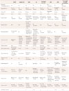
Six research centers with experience and continued interest in the analysis of AD biomarkers in CSF were involved in the creation of this consensus. The opinions of the participants were obtained regarding the individual steps of our own protocols21,28 and those available from international biomarker laboratories or networks, including the Alzheimer's Disease Neuroimaging Initiative (ADNI; http://www.alzforum.org/sites/default/files/protocol_Biofluid_Sample_Collection_Protocol_for_ADNI_0.pdf; http://adni.loni.usc.edu/wp-content/uploads/2008/07/adni2-procedures-manual.pdf), the Alzheimer's Association (AA; http://www.alzforum.org/sites/default/files/protocol_Flow_Chart_for_Lumbar_Puncture_CSF_Processing_0.pdf), Washington University (http://www.alzforum.org/sites/default/files/protocol_CSF_sample_collection.pdf),29 the Alzheimer's Biomarker Standardization Initiative (ABSI),24 the European Network for Biomarkers (ENB),23 and the recent updates of the last two protocols, which focus on the biomarkers of AD and Parkinson's disease (PD)25 (Table 1). If any inconsistencies were found between current and previously used protocols, they were discussed prior to reaching the final consensus. Most of the identified inconsistencies were resolved using a set of higher standards required by more recent protocols, with the hope that the measurements of established and future candidate AD biomarkers in preserved CSF samples would not be influenced significantly.
THE USE OF NEUROIMAGING PRIOR TO LUMBAR PUNCTURE (LP)
When performing a lumbar puncture (LP) to collect CSF, the presence of a high intracranial pressure may result in fatal cerebellar or transtentorial herniation.30 Thus, to exclude the possibility of a mass lesion or elevated intracranial pressure, it is necessary to obtain any brain images at least 1 year prior to the LP. Furthermore, if newly developed headaches or focal neurological signs are present, follow-up neuroimaging should be considered prior to performing an LP. Increased bleeding risk, low platelet count, or the use of anticoagulants would generally prohibit performing an LP, but the continuation of antiplatelet medications is not a contraindication for LP according to the ABSI protocol.24 All procedures need to be performed by experienced physicians.
NECESSITY OF FASTING PRIOR TO LP
The review of the various protocols used by international laboratories revealed controversy regarding the issue of fasting. The ADNI and Washington University require at least 6 hours of fasting (and preferably overnight) prior to an LP, while the ABSI does not. A single Korean study indicated that fasting is mandatory,21 but others indicated that it was not.28,31 Only one study involving a small number of healthy subjects has examined the effects of fasting on Aβ concentrations.32 Even though that study was performed using plasma rather than CSF, the consistency of Aβ levels in CSF (irrespective of fasting) is assumed based on the results of this plasma study.24 However, this evidence is not sufficient to clearly exclude the impact of fasting on the levels of Aβ42, tau proteins, or future candidate biomarkers in CSF. Thus, fasting for a minimum of 6 hours (and preferably overnight) is recommended prior to an LP procedure. Furthermore, if fasting prior to CSF sampling is impossible, the time between the last meal and the LP procedure should be noted.
APPROPRIATE TIMING OF THE LP
Diurnal fluctuations in Aβ42 levels of up to fourfold have been demonstrated,33 highlighting the importance of the time at which the LP required to sample the CSF is performed. However, studies involving elderly populations did not find any significant time-dependent alterations in Aβ42 levels associated with LP.34 The ABSI protocol and its recent updates do not recommend a specific time period for CSF drainage,24,25 but AA protocols suggest an optimal LP procedure time of between 8:00 a.m. and noon (http://www.alzforum.org/sites/default/files/protocol_Flow_Chart_for_Lumbar_Puncture_CSF_Processing_0.pdf), and the ADNI recommends consistency in the timing of LP examinations (http://adni.loni.usc.edu/wp-content/uploads/2008/07/adni2-procedures-manual.pdf). To avoid remarkable diurnal variations in Aβ levels in the CSF, Washington University consistently performs LP procedures at 8:00 a.m.33 Experimental studies using microdialysis procedures in animal models have demonstrated an enhanced clearance of brain metabolites, including Aβ, during sleep,35,36 indicating that diurnal fluctuations in protein levels are an important issue to consider. Thus, it is reasonable to recommend a consistent time range within which the LP procedure should be performed in standardized protocols. Considering the usual schedule of clinical practice in Korea, between 8:00 a.m. and noon has been suggested as a reasonable time period during which to conduct LP procedures. However, exceptions are permitted. Further studies are recommended in order to establish the optimum timing of the LP procedure.
PUNCTURE NEEDLE TYPE FOR LP
The size and type of needle used for LP have not been considered previously as being significant confounding factors when investigating AD biomarker levels in CSF.24 However, it is important to recognize this issue in order to avoid LP-related complications and traumatic punctures.37,38,39 For example, postpuncture headaches and other complications may be highly problematic in elderly patients, especially those with cognitive decline, and the use of an atraumatic needle would probably be beneficial.30 The use of a 22- to 24-G Sporette atraumatic needle is recommended, in agreement with protocols used at most international biomarker laboratories23,24,25 (http://www.alzforum.org/protocols). A conventional cutting-edge needle thinner than 19 G may also be used, but requires a cautious technique, including assuring that the bevel and longitudinal directions of the dura mater fibers are parallel40 and instructing patients to avoid strenuous physical activity for at least 24 hours after the LP procedure.30
CSF DRAINAGE
Natural drainage of the CSF during the LP procedure is recommended. However, if a 24-G atraumatic needle is used, then the CSF sample can be obtained using repetitive 5-mL sterile syringes, as in the ADNI protocol (http://www.alzforum.org/sites/default/files/protocol_Biofluid_Sample_Collection_Protocol_for_ADNI_0.pdf). A sterile polypropylene syringe of the type offered by a standardized manufacturer should be used.
THE NEED FOR ROUTINE ANALYSIS
Cell counts and analyses of total protein and glucose levels using the initially obtained 2-mL CSF sample are required by the protocols of the ADNI, AA, and ENB.23 Although these requirements are not specifically mentioned in other protocols,24,25,29 the routine adoption of this procedure would be beneficial given that blood contamination has been a concern in most studies (as discussed in the following section). Moreover, cell counts and basic chemical profiles should be available as a reference for future investigations. For example, CSF levels of α-synuclein levels, a biomarker for PD, are dramatically influenced by contamination of the CSF by blood.41,42
TRAUMATIC-PUNCTURE ISSUES
Contamination of the CSF by blood is a major concern because it may affect analyses of CSF biomarker concentrations.23,24,25 Although the presence of up to 5,000 red blood cells per microliter in the CSF was found to not significantly alter the levels of Aβ42, which remained within the range of intraindividual variance,32 substantial levels of protein-binding proteins and proteases present in the plasma could potentially affect the measurements of other AD-related biomarkers, such as tau proteins and other metabolites.43 Thus, it is recommended that blood-contaminated CSF be discarded until the sample is clear, as instructed by most international protocols.23,24,25 Furthermore, in the case of a traumatic LP, the immediate centrifugation of CSF is recommended to remove nonvisible blood cells from the fluid.
VOLUME OF COLLECTED CSF
There is controversy regarding the appropriate volume of CSF that should be collected for biomarker analysis. The differences in sampling volumes used among laboratories are worrisome, because it is possible that rostrocaudal gradients influence biomarker concentrations. Accordingly, previous protocols have recommended that the collection of at least 12 mL of CSF is appropriate.23 A small volume of CSF may potentially represent only the contents of the lumbar dural sac, but evaluation of a series of continuous 10-mL CSF samples revealed that Aβ42 levels were not significantly different among the fractions.32 Although no variation in Aβ42 CSF levels has been observed along the spinal cord, other proteins, and especially albumin, neurotransmitters, and apolipoprotein, do vary.44,45,46 Unpublished data using the ABSI protocol24 recommend the collection of at least 1.5 mL of CSF, while the ADNI protocol and Washington University suggest collection volumes of 18 and 30 mL, respectively. The most recent consensus for the appropriate CSF volume to be collected for the preanalytical processing of AD and PD biomarkers recommends a standardized volume of 12 mL.25 Similarly, it is suggested here that the CSF sampling volume be standardized to 10-12 mL for clinical laboratory research.
TYPES OF COLLECTION AND STORAGE TUBES
The types of tube used to collect and store samples for AD biomarker analysis may influence the concentrations of Aβ42 and tau proteins. Polystyrene and glass tubes have been shown to markedly decrease the concentrations of Aβ42, pTau181, and tTau;47 consequently, recent protocols have employed polypropylene tubes.23,24,25,32 However, significant differences in Aβ4248,49,50 and pTau18150 levels have been observed depending on the vendor supplying the polypropylene tubes. These differences demonstrate the necessity of consistency when using these tubes,25,50 especially since numerous commercially available polypropylene tubes are made of copolymers rather than pure polypropylene.24 Accordingly, it is suggested that 15-mL sterile centrifuge tubes with a conical bottom (#352096, BD Falcon, Bedford, MA, USA; or other standardized tubes) should be used as the standard collection tube, as described previously.21 Similarly, the standard storage tubes should be 500-µL sterile polypropylene tubes with screw caps (cat. 72.730.006 or 72.730.005, Sarstedt, Nümbrecht, Germany; or other standardized tubes), as described in the protocols from ADNI and other laboratories.24
NUMBER OF CSF TRANSFERS BETWEEN TUBES
Based on concerns regarding rostrocaudal gradient-dependent variations in AD biomarkers, the Washington University protocol suggests transferring the CSF to new collection tubes following centrifugation to mix the CSF (http://www.alzforum.org/sites/default/files/protocol_CSF_sample_collection.pdf). According to the ADNI protocol, the initially collected CSF should be transferred to two shipping tubes, and a biorepository core laboratory should vortex the CSF thoroughly and aliquot it for storage. However, a mean decrease in Aβ42 levels of 28.6% has been observed following transfer of CSF between tubes, with the most significant change being seen after the initial transfer.51 In the case of tTau, the transfer-induced decrease in concentration is negligible.51 Direct transfer of CSF from the collection tube into individual 0.5-mL frozen tubes is recommended for long-term storage.
STORAGE OF SAMPLES AT ROOM TEMPERATURE PRIOR TO FREEZING
The CSF levels of Aβ42, tau, and pTau are reportedly stable at room temperature for up to 14 days,52 although another study found these protein levels to be constant for only up to 5 days.53 Accordingly, the ABSI protocol for evaluating AD biomarkers permits storage of CSF at room temperature for up to 5 days before its delivery to a core analysis center for freezing.24 However, the stability of other proteins is not certain. The level of a candidate biomarker for synucleinopathy, α-synuclein, is reduced by up to 40% following storage at 4℃ for 4 days,25 and analyses of this protein using surface-enhanced laser desorption/ionization time-of-flight mass spectrometry revealed significant alterations in 6 of the 91 peaks following storage at room temperature for 4 hours.52 Several amino acids and their metabolites are also vulnerable to decay during the time period prior to freezing, even if the delay is only 30-120 min,54 but these changes can be mitigated by the removal of cells via centrifugation. If only Aβ42 and tau proteins are to be analyzed, then delayed freezing may be possible, but the future validation of novel biomarkers for AD diagnosis may require shorter time delays prior to freezing, as suggested by the protocols of the ADNI, Washington University, and ENB,23 as well as the present authors.21 Thus, a delay of no more than 4 hours at room temperature prior to deep freezing is suggested, and it is recommended that the time period before freezing is recorded for each individual sample.
NECESSITY OF CENTRIFUGATION
The ABSI protocol recommends centrifugation only in the case of a traumatic spinal tap,24 while the current ADNI protocol does not include centrifugation. The instability of proteomic analytes due to the time delay before freezing is more profound in noncentrifuged samples.54 Thus, the use of centrifugation to remove nonvisible blood components prior to long-term storage may be reasonable. The most recent protocols for the analysis of AD and PD biomarkers in CSF25 include centrifugation as a mandatory step because α-synuclein levels are susceptible to blood contamination;25,41,42 likewise, the current instructions of the INNOTEST ELISA kit [Fujirebio Europe (previously Innogenetics), Gent, Belgium] specify centrifugation. In addition, different laboratories conduct centrifugation under varying conditions, especially with respect to temperature (room temperature23,24,25 versus 4℃21). Centrifugation does not increase the temperature of the CSF sample.25 Thus, centrifugation at 2,000×g for 10 min at room temperature is reasonable based on previous protocols23,25 and the instructions provided by the kit manufacturer.
ALIQUOT VOLUME AND LABELING OF THE FROZEN TUBES
Concerns regarding freeze evaporation and surface adsorption have been raised; as a result, some protocols specify the aliquot volume occupy at least 50%24 or 75%23,25 of the tube volume, and the use of a screw cap (http://www.alzforum.org/sites/default/files/protocol_CSF_sample_collection.pdf). Moreover, additional confounding factors related to aliquot volume have been identified recently. Levels of Aβ42, but not tTau or pTau, change significantly depending on the storage volume of the CSF samples.55 The high hydrophobicity of Aβ42 may increase surface adsorption to the vessel wall, but addition of the Tween-20 detergent to the initial CSF samples prior to aliquoting diminishes the adsorption of Aβ42 into polypropylene tubes.55 However, this manipulation step has yet to be adopted by established protocols. Thus, it is important to standardize the aliquot volume and the type of tubes used for long-term freezing. The use of 500-µL polypropylene tubes with screw caps (500 µL; cat. 72.730.006 or 72.730.005, Sarstedt) to aliquot 400 µL of CSF and immediate freezing of the samples at -80℃ are recommended. According to the INNOTEST kit, the total volume of CSF per aliquot required for measurements of Aβ42, t-Tau, and p-Tau (with duplicates) is 350 µL. Thus, a fresh aliquot volume of 400 µL is sufficient for the measurement of these three biomarkers.
Subsequently, written labels that include the coded sample numbers and the serial aliquot number of the total numbers of aliquot for each CSF sample (e.g., 3/20, the 3rd of 20 aliquots) are recommended on the surface of frozen tubes. Water-resistant materials should be used for labeling.
NUMBER OF PERMITTED FREEZE-THAW CYCLES PRIOR TO ANALYSIS
The importance of minimizing the number of freeze-thaw cycles is clear, and accordingly most protocols limit the number of freeze-thaw cycles to one or two23,24,25 (http://www.alzforum.org/protocols). Nonetheless, there is controversy concerning the extent of the susceptibility of the Aβ42, pTau, and tau proteins in CSF to freeze-thaw cycle repetitions. A reduction in Aβ42 levels has been observed in some studies even after the first freeze-thaw cycle,56,57 but not in others.32,58 Levels of tTau and pTau, but not Aβ42, were altered after two and three freeze-thaw cycles, respectively,52 but another study found that the levels of Aβ42, pTau, and tTau did not vary significantly when freeze-thawing was repeated up to three times.53 Instability of other CSF-related proteins has been observed in studies using proteomic methods,52 raising concerns about potential unidentified effects on AD biomarkers. Thus, subjecting the samples to only one freeze-thaw cycle is recommended.
CONCLUDING REMARKS
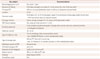
There is a better understanding of the biological mechanisms underlying the clinical manifestations of AD than perhaps those underlying other neurodegenerative disorders, and AD-related biomarkers therefore have implications for clinical practice. The study of these markers in CSF has thus become more than just of academic interest. The general acceptance of Aβ42, pTau, and tTau levels in CSF as biomarkers for the early diagnosis of AD was evident after their inclusion in the new diagnostic criteria for AD established by the National Institute on Aging and the Alzheimer's Association.4 With regard to the development of disease-modifying therapies in the future, the early diagnosis of AD is of prime importance for therapeutic interventions. Even before that time, the precise diagnosis of AD during the early stages is beneficial for initiating aggressive and appropriate available treatment. Currently, the measurements of pTau181 in CSF using ELISA have been accepted as a new health technology in Korea (http://neca.re.kr/nHTA/index.jsp). Although the diagnostic cutoff values for Aβ42, pTau181, and tTau in CSF for the diagnosis of AD have been established in one Korean laboratory,21 the direct application of these values by other institutions should be approached with caution. There is distinct interlaboratory variability in the measurement of AD biomarkers internationally among biomarker laboratories, especially for Aβ42.22 Standardization of the preanalytical sampling and handling protocols is necessary to ameliorate this variability (Table 2), and its importance cannot be overemphasized. Thus, in the present article we have made a concerted effort to review, assess, and suggest the most reliable procedures for avoiding any disturbances during the preanalytic process. The analysis of AD biomarkers in CSF will hopefully be conducted under the supervision of a quality control program according to this cooperative protocol, to achieve the ultimate goal of a qualified and valid AD biomarker in CSF that can be assessed with high reliability in Korea.
 XML Download