

 PDF
PDF ePub
ePub Citation
 Print
Print


Introduction
Metamorphopsia includes a broad spectrum of visual perceptual distortions, such as alteration of perceived object size or, rarely, altered perception of faces, termed prosopometamorphopsia. Prosopagnosia, on the other hand, is a neurological deficit characterized by an inability to recognize faces despite intact intellectual and cognitive function. The fusiform face area (FFA) of the brain is known to play a key role in face perception. Several studies have shown that the perception of facial configuration is impaired in patients with prosopagnosia whose lesions involve the right fusiform gyrus.1 We report herein a patient who complained of metamorphopsia restricted to the center of the face, and particularly the lower part of the face (nose and mouth), following right medial temporooccipital lobe infarction that included the FFA.
Case Report
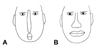
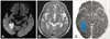
A 75-year-old right-handed woman was admitted for the sudden onset of nausea, dizziness, and blurred vision. She had suffered from congestive heart failure, hypertension, and atrial fibrillation for more than 20 years. She had no previous history of hyperlipidemia, diabetes mellitus, or any ophthalmic disease. Routine biochemistry laboratory parameters and the full blood cell count were within normal limits. She complained of dimmed vision, and she described the central part of faces, particularly the nose and mouth, as being out of shape. Regardless of whether she looked at a familiar or an unknown person, she claimed that "the nose looks very narrow and lengthened toward the mouth, which looks small and round in shape" (Fig. 1A). According to her description, she appeared to see faces as if viewed through a convex lens. She had no prosopagnosia; when presented with images of ten famous Korean faces (e.g., actors, singers, or the president), she was able to readily identify all of them and she could correctly recognize images of objects. She had no impairment in her visuoperceptual performances (describing a complex scene, drawing figures, reading, and writing) or in color perception. No hemineglect was found, as assessed using a line bisection or star-cancellation task and copy-drawing a flower and a clock. Other components of the neurologic examination were normal, including cranial nerves, motor and sensory functions, cerebellar function, reflexes, and gait. The rest of the physical examination was unremarkable. In particular, she had no cognitive or psychiatric impairment; the Mini-Mental State Examination and Seoul Neuropsychological Screening Battery were performed at her bedside. She copied the Rey-Osterrieth figure accurately, but had trouble recalling it. While she also had verbal memory difficulties, this was considered normal when allowing for her age. Left homonymous quadrantanopia was detected by Humphrey automated perimetry. Diffusion-weighted MRI and T2-weighted brain MRI revealed an infarction in the right medial temporooccipital lobe that included the parahippocampal gyrus (Fig. 2). No evidence of stenosis or occlusion of the intracranial arteries was observed on MR angiography. The patient was treated with intravenous heparin infusion and coumadization over 10 days; at the time of discharge she had recovered to the extent of near normalcy in describing faces (Fig. 1B).
Discussion
Face perception may be the most highly developed visual skill in humans. Merely looking at somebody's face allows us to determine the age, sex, and attractiveness of the person, and further, to estimate his or her mood and friendly or hostile attitudes. Although each human face consists of the same components, most of us have the unique maximal capacity for memorizing instances of these social stimuli.2 The existence of a specialized neural system for face perception in the brain was suggested by the observation of patients with focal brain damage who had a prosopagnosia. However, the type of processing required for face recognition in general is an area of ongoing research.
In terms of classical face recognition models,3 there are several modules in the brain that independently process information about human faces. It has been reported that to recognize a face, the brain must process information related to the changeable face aspects-such as facial expression and eye gaze direction-separately from features that are invariant. This is why a small facial modification will not be misinterpreted as a change in face identity. Such processes are controlled by the so-called core system, which comprises three bilateral regions: the inferior occipital gyrus, the lateral fusiform gyrus, and the superior temporal sulcus. Other parts of the brain also participate in face perception as they are recruited to process the significance of information gleaned from the face: "the extended system" (including the amygdala and insula), where facial expressions are processed, the intraparietal sulcus, the auditory cortex, and the anterior temporal area. Therefore, it is likely that the processing of facial information requires the integration of activity across a network of cortical regions, not just within a single region.
Studies of prosopagnosia patients have confirmed the existence of an FFA located in the midsection of the fusiform gyrus.4 Many functional imaging studies of face perception have found that the FFA exhibited a stronger response to faces than to assorted common objects.5 Although the FFA also responds significantly to other objects, it is commonly believed to be a face-selective cortical region dedicated to the visual analysis of face stimuli. But the question still remains as to whether the FFA alone is sufficient, or even necessary, for face perception. Some prosopagnosia patients, despite their profound inability to recognize faces, exhibit normal patterns of activation in the FFA, suggesting that activation in the region alone is not sufficient for face recognition, which likely depends on integration across cortical regions.6
In this paper we describe a patient with a subjective complaint of prosopometamorphopsia (restricted to the lower half of face) with right medial temporooccipital lobe lesions involving the parahippocampal gyrus. The parahippocampal gyrus is considered to be the complement of the FFA. This patient did not exhibit prosopagnosic disturbances, but complained about altered perception of faces.
Prosopometamorphopsia can involve the whole face, but it can also affect only one side of the face, usually after right hemisphere damage. There are several reports of unilateral prosopometamorphopsia for the right half of faces after right posterior temporal damage,7 but unilateral prosopometamorphopsia after left hemisphere lesion has rarely been described. However, to our knowledge there has been no report of prosopometamorphopsia restricted to the nose and mouth, either involving whole or unilateral face perception.
In the light of recent empirical evidence, face perception is thought to be mediated by the distribution of central neural systems, including all regions of the core and extended systems, of which the main entry node is the lateral fusiform gyrus. Face-responsive regions in occipital areas of each hemisphere perceive contralateral sides of the face; thereafter the facial information is directed toward the FFA of the right hemisphere, where information from each side is integrated. Therefore, we speculate that any injury on this pathway could induce prosopometamorphopsia involving whole or unilateral face perception, or very rarely, such as in our case, distortion restricted to the central area of the face. Furthermore, we hypothesize that there could be topographical correspondences between facial structures and the FFA. Given that face perception is one of the most highly developed visual skills, the FFA is likely to include an area that is specifically responsible for recognizing faces as topographical patterns. However, the analysis of our prosopometamorphopsia patient was based mainly upon the patient's subjective complaints, thus preventing any attempt to elucidate its mechanisms. Further accumulation of similar cases is needed to collect evidence to gain a better understanding of this type of face metamorphopsia.
 XML Download