

 PDF
PDF ePub
ePub Citation
 Print
Print


Introduction
Dyslipidemia contributes to the development of atherosclerosis,1-3 and there is accumulating evidence that atherosclerosis is promoted by remnant lipoproteins (RLPs).4-13 These are products of partially catabolized chylomicrons and very-low-density lipoproteins, from which some triglycerides (TGs) have been removed by the action of lipoprotein lipase and, to a lesser extent, by hepatic lipase. These particles have reduced TGs, but are enriched with cholesterol and apolipoprotein (apo)E; they are smaller and denser than the parent particles, and are believed to be more strongly atherogenic than larger TG-rich lipoproteins.4-9
Elevated RLP cholesterol (RLP-C) levels were reported to be associated with endothelial dysfunction, an early marker for atherosclerotic disease.10 The Framingham Heart Study found that an increase in RLP-C levels was a significant risk factor for coronary artery disease in women.11 In another study with a 3-year follow-up period, the incidence of cardiovascular events in the high RLP-C group (RLP-C >5.1 mg/dL) was higher than that in the low RLP-C group (RLP-C ≤3.3 mg/dL).12 As a result, in 2000 the United States Food and Drug Administration recognized that an increase of RLP-C was a risk factor for coronary heart disease.13
Nakamura et al.14 recently reported that high levels of RLPs predict ischemic stroke in patients with metabolic syndrome and mild carotid atherosclerosis. However, the patient population in that study comprised a selected subgroup of patients who were admitted for cardiac catheterization for chest pain or electrocardiographic ischemic changes. In the present study of consecutive patients with ischemic stroke, we aimed to add new information regarding the association between RLP-C and ischemic stroke and, given that stroke is a heterogeneous disease with more than 150 known causes,15 to identify the correlations of RLP-C levels with various stroke subtypes.
Methods
Study patients
This study was approved by the institutional review board of the Dongguk University Ilsan Hospital (DUIH), Korea. From February to September 2007, 198 consecutive patients who were admitted to the DUIH with acute ischemic stroke (≤1 week) were prospectively screened for enrollment. Acute ischemic lesions were assessed by diffusion-weighted imaging with the apparent diffusion coefficient. We excluded three patients with incomplete evaluations due to severely impaired consciousness or early discharge, four patients with transient ischemic attack without visible lesions on diffusion-weighted imaging, and six patients with cerebral infarction due to venous thrombosis, arterial dissection, or moyamoya disease. Sixteen patients were excluded because their lipid profiles included only total cholesterol. We excluded a further 27 patients who did not fast for at least 9-12 hours before the measurement of lipid or RLP-C levels. Thus, a total of 142 patients (90 men and 52 women; age, 65.2±12.8 years, mean±SD) were entered into the study. All patients underwent systemic investigations including assessment of medication history, magnetic resonance imaging (MRI) with MR angiography, carotid duplex ultrasonography, transthoracic echocardiography, 24-h Holter monitoring, and other routine admission laboratory tests. We prospectively collected demographic data, prior medication history, and the presence of vascular risk factors including hypertension, diabetes mellitus, heart disease, previous stroke, and smoking.
Assessment of stroke risk factors
Hypertension was considered to be present if a subject had one of the following conditions: repeated blood pressure readings above 140/90 mm Hg at 1-2 weeks after stroke onset, a history of hypertension, or use of antihypertensives. Diabetes mellitus was defined as a glycated hemoglobin (HbA1C) level of ≥6.5%, a history of diabetes mellitus, or use of diabetic medication. Smokers included current smokers, who smoked daily or only occasionally. Heart disease as a potential source of cerebral embolism16 was considered to be present if a subject had a left atrial or ventricular thrombus, atrial fibrillation or sick sinus syndrome, recent myocardial infarction within 1 month, rheumatoid mitral or aortic valve disease, bioprosthetic or mechanical heart valves, chronic myocardial infarction with a low ejection fraction of <28%, symptomatic congestive heart failure with an ejection fraction of <30%, dilated cardiomyopathy, aneurysms or akinetic segments of the left ventricular wall, or nonbacterial thrombotic endocarditis. Electrocardiography, transthoracic echocardiography, and 24-h Holter monitoring were performed to identify these cardioembolic sources. Transesophageal echocardiography or a transcranial Doppler bubble study was also performed in selected patients.
Blood samples were taken in the morning after an overnight fast of 9-12 h (12.0±3.2 h in the stroke group and 12.0±0.1 h in the control group). In the stroke subjects, the median sampling time from stroke onset was 39.8 h. Total cholesterol, TG, high-density lipoprotein cholesterol, and low-density lipoprotein cholesterol levels were assayed by enzymatic techniques. RLP-C was measured in triplicate after isolation, as reported previously,17 by using an immunoaffinity gel containing monoclonal antibodies to human apoB-100 and apoA-I (Jimro-II RLP-C kit, Otsuka, Japan). Mean values were used for the analysis. Serum RLP-C levels were considered to be high when they were within the highest quartile (>75th percentile)18 of the values of all of the study participants.
Stroke subtype analysis
Subtypes of index stroke were determined by the consensus of three neurologists (M.G.J., S.W.J., and D.E.K.) as follows: large artery atherosclerosis (LAA), small vessel occlusion, cardioembolism (CE), and ischemic stroke of undetermined etiology, as described in the Trial of Org 10172 in Acute Stroke Treatment study.16
Control subjects
As a control group, we chose 88 consecutive subjects (34 men and 54 women; age, 54.4±9.3 years) 1) who visited the DUIH Healthcare Center for health screening during the same study period, 2) who did not have a history of stroke, transient ischemic attack, myocardial infarction, or angina, and 3) whose lipid profiles and RLP-C were measured after 9-12 h of fasting.
Statistical analyses
Data are presented as mean±SD values. Comparisons of continuous variables between groups were performed using Student's t-test, one-way analysis of variance (ANOVA) with Dunnett's post hoc test, or Kruskal-Wallis ANOVA with post hoc Mann-Whitney test. The chi-square test or Fisher's exact test was used to compare proportions between groups. Bivariate correlations were calculated to quantify the relationships between pairs of variables. Multivariable regression analyses were conducted to determine if high RLP-C level was an independent risk factor for ischemic stroke or an independent predictor for LAA stroke after adjustment for known vascular risk factors. All statistical analyses were conducted using the SPSS software package (SPSS 18.0, Chicago, IL, USA). A value of p<0.05 was considered statistically significant.
Results
Univariable analysis showed that RLP-C is a risk factor for ischemic stroke
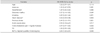
Table 1 summarizes the demographic and clinical features of the study subjects. Compared with the control group, the stroke group had higher mean values or prevalences of the vascular risk factors including age, male sex, hypertension, diabetes, heart disease, previous stroke, smoking, and TG level (all p<0.05). However, the serum levels of total cholesterol, low-density lipoprotein cholesterol, and high-density lipoprotein cholesterol were higher in the control group than in the stroke group (all p<0.05). In line with our hypothesis, the univariable analysis showed that RLP-C levels were higher in the stroke group than in the control group (p=0.043). Similarly, when RLP-C levels were dichotomized at 5.56 mg/dL (which was the 75th percentile boundary), a significantly higher proportion of the stroke group had high RLP-C levels (31.0% vs. 14.8%, p=0.006).
Multivariable analyses showed that a high RLP-C may be an independent risk factor for ischemic stroke
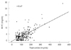
The association between RLP-C and stroke could be due to various confounding factors; therefore, logistic regression analyses were conducted, where those variables with p<0.15 in the univariable analyses were included. The continuous variable "RLP-C" did not satisfy the linearity assumption of logistic regression; therefore, the dichotomized variable "high RLP-C" was entered into the model. After adjusting for age, sex, hypertension, diabetes mellitus, smoking, heart disease, previous stroke, total cholesterol, and statin medication, the odds ratio for the association of high RLP-C with stroke was 2.54 (p=0.045) (Table 2). When TG was also entered into the model, a high RLP-C was not significantly associated with stroke (p=0.24). TG was also not significantly associated with stroke (p=0.21). The scatterplot and Pearson correlation analysis (Fig. 1) revealed that there was a linear relationship between RLP-C and TG (p<0.01, r=0.67).
RLP-C is an independent predictor for LAA stroke versus the other stroke subtypes
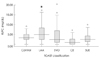
As indicated in Table 3, which presents the risk factors in the stroke subgroups based on the Trial of Org 10172 in Acute Stroke Treatment classification, the RLP-C level was highest in the LAA subgroup (5.73±3.94 mg/dL) and lowest in the CE subgroup (1.76±2.23 mg/dL). As illustrated in the box plots and demonstrated by the Kruskal-Wallis ANOVA with Mann-Whitney post hoc tests (Fig. 2), the RLP-C level was significantly higher in the LAA subgroup than in the subgroups (all p<0.05). Similarly, the RLP-C level was more frequently in the highest quartile in the LAA subgroup than in the other groups (43.8% vs. 10.5-31.5%). Other stroke risk factors and traditional lipid profiles, with the exception of TG levels, did not differ significantly among the subtypes. Unlike for the RLP-C levels, the serum TG levels of the LAA subgroup were not higher than those of the other subgroups except the CE subgroup. Table 4 presents the independent factors associated with LAA vs. other stroke subtypes. After adjusting for TG as well as age, sex, hypertension, diabetes mellitus, smoking, heart disease, previous stroke, total cholesterol, and statin medication, a high RLP-C was the only significant predictor that was positively associated with the LAA stroke vs. the other stroke subtypes (p=0.045). Heart disease and diabetes were negatively associated with LAA stroke (p=0.005 and 0.049, respectively).
Discussion
In this study we observed a significant association between higher RLC-C levels and ischemic stroke in general, with a particular association between RLP-C and LAA stroke. A high RLP-C was four times more prevalent in patients with LAA stroke (44%) than in the CE subgroup (11%). In multivariable analysis, a high RLP-C was linked with ischemic stroke after adjusting for prior statin medication and known vascular risk factors. TG levels were correlated with RLP-C levels, and after adjusting for TG as well as other risk factors, a high RLP-C was the only significant predictor that was positively associated with LAA stroke. This suggests a separate significance for RLP-C levels independent of TG levels. Together these findings indicate the potential utility of examining RLP-C levels for stroke risk assessment.
RLP-C has not been widely studied due to a lack of routine laboratory assays; until the recent development of a simple and reliable technique for measurements based on an immunoseparation method,17 it has been difficult to assay levels of RLP-C. In the field of stroke, only one recent study14 has demonstrated that high RLP-C levels (≥5.8 mg/dL) were related to echolucent carotid plaques and shown to be an independent risk factor for future ischemic strokes in a subgroup of patients admitted for cardiac catheterization, who had metabolic syndrome and evidence of carotid plaques producing stenosis of less than 50%. The present cross-sectional study found that an ischemic stroke was about 2.5 times more likely in subjects with RLP-C levels higher than 5.56 mg/dL than in the controls. This supports the notion that RLP-C may be related to the genesis of cerebral vascular events,14 which could be mediated by the proinflammatory and proatherothrombogenic effects of RLP-C:18-21 inhibition of nitric-oxide-mediated arterial dilation, and up-regulation of endothelial expression of intracellular adhesion molecule-1 and vascular cell adhesion molecule-1.
The finding that high RLP-C levels were more frequently encountered in patients with LAA stroke supports a role for RLP-C in proatherosclerotic endothelial dysfunction,18-20 which would be expected to play a larger role in this group than in patients with embolic vascular occlusions. It is possible that future studies will allow RLP-C levels to be used for determining the contribution of endothelial dysfunction to stroke, particularly when it is not clear if in situ atherosclerosis or embolism was responsible for a lesion. This may have important clinical implications because the management and prognosis of stroke patients are influenced by the ischemic stroke subtype.
The mean RLP-C level of our control group was 3.62 mg/dL, which is similar to the values reported for Japanese control subjects (3.3 or 3.7 mg/dL).19-23 The mean RLP-C level of the stroke group was 4.5 mg/dL, while the LAA subgroup had the highest mean value, at 5.73 mg/dL. These findings are in line with previous reports that patients with RLP-C levels higher than 4.7-5.1 mg/dL are at increased risk of cardiovascular events.12 The stroke patients in our CE subgroup had the lowest mean RLP-C level, 1.84 mg/dL, which was lower than the mean value for the control group. The patients in the CE subgroup had atrial fibrillation or ventricular akinesia as a cause of the index stroke; they did not have significant atherosclerotic narrowing of the large artery supplying the infarct territory. These data indicate that high RLP-C levels are associated with atherosclerotic stroke, not cardioembolic stroke.
There are several mechanisms by which RLPs can lead to endothelial dysfunction and stroke. The findings of a previous in vitro experiment suggested that macrophage foam cells in atherosclerotic plaques, which could secrete inflammatory proteases in or around the complicated areas of the human atheromata,24 are derived from the cellular uptake of chylomicron remnants.25 In addition, high levels of RLPs were reported to cause endothelial vasomotor dysfunction in human coronary arteries.18 RLP-C was also shown to up-regulate the expressions of both intracellular adhesion molecule-1 and vascular cell adhesion molecule-1 in cultured human endothelial cells,19 which could promote atherosclerosis.20,21 Moreover, high levels of RLP-C may contribute to the development and thrombotic complications of atherosclerosis via the combined effects of up-regulation of endothelium-derived proatherothrombogenic molecules and enhanced platelet reactivity.22 Our finding that high RLP-C levels were associated with LAA stroke is consistent with and provides a potential explanation for the findings of the aforementioned studies.
Our study was subject to some limitations. First, the study had a cross-sectional design and therefore did not allow us to make causal inferences. Second, the control group was not fully representative of the source population from which the stroke patients were selected, although all the subjects were consecutively enrolled from the same hospital during the same study period, and multivariable analyses were used to adjust for potential confounders.
In conclusion, we have demonstrated an association between elevated levels of RLP-C and LAA stroke.

 XML Download