

 PDF
PDF ePub
ePub Citation
 Print
Print


Introduction
A resurgence of pertussis has been observed in a number of countries with highly vaccinated populations, and pertussis has become the most prevalent vaccine-preventable disease in industrialized countries [1]. In Korea, the diphtheria-tetanus-acellular pertussis (DTaP) vaccination rate is above 94% [2]; however, pertussis has been confirmed since 2000 in adolescents and adults [3]. Furthermore, a small outbreak of pertussis was reported among young children in 2009 [3]. Recently, pertussis has been increasing among adolescents and adults in many countries with high vaccine coverage rates because of waning acquired immunity after DTaP vaccination [4]. In addition, there have been several reports abroad of mixed infections of pertussis and other respiratory pathogens [567]; however, no such cases have been reported in Korea. We report a case of coexisting pertussis with mycoplasma pneumonia in a child who presented with prolonged cough.
Case Report
A 10-year-old girl was admitted to our hospital with complaints of 10 days of cough and sputum. She had been treated at a private hospital with a diagnosis of acute bronchitis; however, her fever continued. She also had paroxysmal cough with post-tussive emesis since six days before hospitalization. She did not have a previous history of asthma or hospitalization and had received five doses of DTaP vaccination.
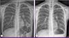
On admission, the patient presented with blood pressure of 100/80 mmHg, heart rate of 102 beats/minute, respiratory rate of 20 breaths/minute, body temperature of 38.2℃, and alert mental status. Physical examination revealed no abnormal signs except for crackles on both lung fields. Laboratory tests showed leukocyte count of 3,960/mm3 (neutrophils 58.8%, lymphocytes 27.5%), hemoglobin 11.0 g/dL, platelet count 198,000/mm3, erythrocyte sedimentation rate of 50 mm/hour, and C-reactive protein 1.69 mg/dL. Blood chemistry and urine testing were unremarkable. A chest X-ray showed diffuse pneumonic infiltrations in the left lower lung field (Fig. 1A). Empirical roxithromycin therapy (2.5 mg/kg twice a day) was initiated on admission because she was diagnosed with atypical pneumonia and M. pneumoniae is epidemiologically the most common cause of atypical pneumonia in Korea [8].
On hospital day 3, blood and urine cultures gathered on admission revealed no pathogenic microorganisms, and the tuberculin skin test was negative. The multiplex polymerase chain reaction (PCR) for respiratory viruses including respiratory syncytial virus, rhinovirus, human metapneumovirus, parainfluenza virus, influenza A/B virus, and adenovirus (AdvanSure™ RV real-time PCR, LG Life Sciences Ltd., Seoul, Korea) which was performed on admission was negative. The serologic tests for M. pneumoniae was performed using commercial enzyme-linked immunosorbent assay (ELISA) kits (Chorus M. pneumoniae IgG and Chorus M. pneumoniae IgM, DIESSE Diagnostica senese S.p.A., Siena, Italy), and qualitative IgM antibody and semi-quantitative IgG antibody (titer = 95 AU/mL) against M. pneumoniae were positive. The cold agglutinin titer was 1:64. She was diagnosed with mycoplasma pneumonia based on the elevated cold agglutinin titer and positive IgM and IgG against M. pneumoniae. Her paroxysmal cough did not improve during hospitalization, and a PCR for pertussis using her nasal aspirates was conducted on hospital day 4. The in-house PCR assay (forward primer: 5'-GATTCAATAGGTTGTATGCATGGTT-3', reverse primer : 5'-TTCAGGCACACAAACTTGATGGGCG-3') was performed based on the guidance of the European Centre for Disease Prevention and Control [9]. The PCR for pertussis was positive, and eventually she was diagnosed with coexisting pertussis with mycoplasma pneumonia. Her family members were screened for pertussis with a commercial ELISA kit (Bordetella PT IgG ELISA, IBL International GmbH, Hamburg, Germany). Although none of her family members manifested respiratory symptoms, her father showed an elevated anti-pertussis toxin (PT) IgG titer of 106 IU/mL; the anti-PT IgG titer higher than 100 IU/mL which indicates recent pertussis infection. Other family members were having lower than 100 IU/mL anti-PT IgG titers. The chemoprophylaxis for family members was recommended; however, they refused to follow our suggested guides.
On hospital day 7, the patient's symptoms began to improve and crackles on both lung fields disappeared. A repeat chest X-ray showed improvement of pneumonic infiltrations (Fig. 1B). On hospital day 10, she was discharged from the hospital, but continued roxithromycin therapy. She re-visited the outpatient clinic four days after discharge, and the antibiotic therapy was completed at that time. Repeat laboratory tests revealed that the cold agglutinin titer was 1:128, and IgM and IgG (titer >100 AU/mL) antibodies were still positive.
Discussion
In spite of DTaP vaccinations, pertussis has steadily increased in many countries during the last two decades with prominent incidence in adolescents and adults [11011]. Pertussis tends to present with no or atypical symptoms in adolescents and adults; therefore, the appropriated diagnosis and treatment can be delayed. Additionally, such undiagnosed pertussis patients have become an important source of pertussis transmission to other household members, particularly infants and young children who are not fully immunized [4]. In Korea, 66 pertussis patients were reported in 2009, and 37% of their family members showed positive results of the pertussis diagnosis test, meaning a probable risk of pertussis outbreak existed [12].
Our patient was initially tested for M. pneumoniae and respiratory viruses based on her respiratory symptoms and chest X-ray findings, and she was diagnosed with mycoplasma pneumonia. Because M. pneumoniae grows slowly in culture, serologic tests have been the most commonly used diagnostic modality instead of culture, thereby, the usefulness of PCR have been reported nowadays [1314]. As for the serologic methods, the complement fixation test was previously the most used method; however, the enzyme immunoassays using commercial kits are widely used recently [1314]. Serum cold agglutinin titers have been used for the diagnosis of mycoplasma infection for a long time, although it is not specific for M. pneumoniae [1315]. The elevated cold agglutinin titers can also be observed in other viral infections, such as adenovirus, influenza virus, and Epstein-Barr virus; however, they are detected in low cold agglutinin titers ranging from 1:2 to 1:32 [15]. The mycoplasma infections should be considered if cold agglutinin titers are 1:64 or higher [15]. Because IgM antibody may persist for several months to years after mycoplasma infection, the fourfold increase in IgG titers of paired sera is necessary for a diagnosis of acute mycoplasma infection, and acute mycoplasma infection should be questioned when the IgM antibody is positive with a negative cold agglutinin test result [13]. Furthermore, positive PCR results cannot be excluded of simple colonization. As a result, some experts recommend using both PCR and serology results at the same time in diagnosing acute mycoplasma infection [1516]. In our patient, both IgM and IgG antibodies against M. pneumoniae were initially positive which led us to consider both recent and remote mycoplasma infections. Eventually, we diagnosed her with recent mycoplasma infection with a high cold agglutinin titer of 1:64. After then, the patient complained of paroxysmal cough and subsequent emesis lasting for more than a week, and therefore an additional pertussis test was performed. The patient did not present with typical whooping cough, and laboratory tests did not show absolute lymphocytosis. When children and adults with partial immunity against pertussis are infected, typical pertussis symptoms and signs, such as whooping or absolute lymphocytosis, are often replaced with nonspecific symptoms, resulting in under-diagnosis [1718]. These undiagnosed pertussis-infected patients may spread the infection, as was noted in this case. The patient's father showed an elevated anti-PT IgG titer higher than 100 IU/mL, suggesting recent pertussis infection.
In accordance with the increasing incidence of pertussis infection, various diagnostic methods for pertussis using pertussis antigens such as PT and PCR for pertussis have been developed. However, diagnostic tests for pertussis are seldom performed because of the relatively rare occurrence of pertussis compared with respiratory viruses and mycoplasma infection. In addition, clinicians may not perform pertussis tests if other respiratory pathogens are identified in patients with respiratory symptoms, despite the possibility of co-infection. Several reports noted that mixed infection of pertussis and adenovirus was common, and symptoms were more severe in younger patients compared with older ones [1920]. Another study of 135 children with chronic cough of one to six weeks reported that the most common causes of chronic cough were rhinovirus followed by pertussis and respiratory syncytial virus [6]. In that study, the most common combination of mixed infection among respiratory pathogens was pertussis and rhinovirus [6]. In another study, 28% of serologically confirmed pertussis patients were identified to have mixed infection with respiratory viruses or M. pneumoniae [5].
In conclusion, the present case is the first report in Korea of a mixed infection of Bordetella pertussis and M. pneumoniae in a child who completed DTaP vaccination. If a respiratory virus or M. pneumoniae are identified in a patient and the patient's symptoms improve, the effort to find other pathogens may be neglected. However, pertussis may co-infect such patients and result in atypical symptoms depending on the patient's immunity against pertussis. If untreated, these patients may spread pertussis to the broader population. A test for pertussis is recommended in patients who present with prolonged cough as a chief complaint in order to reduce the potential for spreading pertussis.
 XML Download