

 PDF
PDF ePub
ePub Citation
 Print
Print


Introduction
Although Corynebacterium species are ubiquitous gram-positive pleomorphic aerobes that colonize the skin and mucous membranes in humans, they rarely account for clinical infections [1]. Until recently, the pathogenic potential of coryneform bacteria has been underestimated and has been overlooked as a mere skin contamination. However, recent reports show that C. macginleyi isolated from the ocular sites can be the cause of conjunctivitis, keratitis, and endophthalmitis [2-5]. In addition, reports that describe Corynebacterium species as the causative agents for significant and life-threatening infections such as pneumonia, vertebral osteomyelitis, bacteremia, device related infections, endocarditis, and abscesses in immunocompromised patients are increasingly presented [6-8]. Herein we present for the first time the possible involvement of a C. macginleyi strain as the causative agent of pneumonia in a human immunodeficiency virus (HIV) patient.
Case report
A 42-year-old homosexual man was hospitalized with a 1-month history of fever. He complained of dry cough, weakness, fatigue, and loss of appetite which developed 12 weeks previously. On admission, his body temperature was 38℃ and respiratory rate was 33/min. Pulmonary crackles were heard in both lower lung fields. The patient's PaO2 was 47.4 mmHg and O2 saturation was 88.9% on room air. Leukocyte count was 2,950/mm3 (58% neutrophils and 26.1% lymphocytes). Enzyme-linked immunosorbent assay for HIV was positive and this was confirmed by Western blot analysis. CD4 lymphocyte count was 14/mm3 and viral load was 13,000 copies/mL. The chest radiograph revealed ill defined ground glass opacities in both lungs with central and upper lobe predominance (Fig. 1). He was treated with trimethoprim-sulfamethoxazole (TMP/SMX), steroid, lopinavir/ritonavir, lamivudine, and zidovudine. Methenamine silver stain of bronchoalveolar lavage fluid was negative and the culture revealed no organisms. His general condition improved and fever abated on day 3 after the initiation of treatment. Therefore, we considered P. jirovecii to be the causative agent of pneumonia. The patient was then discharged after showing clinical improvement with three weeks of therapy.
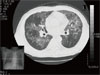
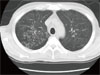
Five weeks later, he was admitted again with a 10-day history of cough and mucopurulent sputum. On admission, his vital signs were stable; body temperature was 37.4℃ and respiratory rate was 18/min. Laboratory results were as follows: PaO2, 82.1 mmHg and O2 saturation, 97% on room air; leukocyte count, 6,940/mm3 (78% neutrophils and 13.8% lymphocytes), erythrocyte sedimentation rate, 90 mm/h; and CD4 lymphocyte count, 101/mm3. Since crackles were heard on both lower lung fields, chest CT scan was taken and it showed multifocal clusters of ill defined small centrilobular nodular opacities with bronchial wall thickening and linear opacities in both lungs (Fig. 2). Treatment was initiated with TMP/SMX and IV cefuroxime (750 mg q 8 h) for presumed Pneumocystis jirovecii pneumonia with or without combined community acquired pneumonia. Bronchoscopy revealed diffuse acute inflammatory changes of the bronchial mucosa with abundant purulent bronchial secretions from both bronchi but methenamine silver stain for P. jirovecii was negative. However, the bronchial washing fluid culture yielded > 100,000 colonies/mL of C. macginleyi that was susceptible to vancomycin, gentamicin, and tobramycin, but resistant to penicillin, oxacillin, cephalothin, ciprofloxacin, and TMP/SMX. On day 6 after the initiation of antibiotic treatment, the patient developed fever and complained of shortness of breath. We therefore considered corynebacteria to be the causative agent of pneumonia and thus vancomycin was administered. He fully recovered after receiving a 14-day course of antibiotic therapy with vancomycin monotherapy.
Discussion
Pulmonary infections are a leading cause of morbidity and mortality in persons with HIV infection [9, 10]. Following the introduction of highly active antiretroviral therapy and TMP/SMX chemoprophylaxis, the relative incidence of HIV-associated pneumonia has changed. Whereas the incidence of P. jirovecii pneumonia and tuberculosis declined, bacterial pneumonia have become the most frequently encountered HIV-associated opportunistic respiratory infections [11, 12]. Common respiratory tract bacterial pathogens (i.e., Streptococcus pneumoniae and Haemophilus influenzae) and less pathogenic bacteria, such as the coryneform bacterium, have been implicated as the causative agents [10-12].
Species of the genus Corynebacterium are widely distributed in the environment as normal inhabitants of soil and water. They are gram-positive, non-acid-fast, aerobic or facultatively anaerobic, asporogenous rods [1]. In the hospital setting, they may be cultured from the hospital environment, including surfaces of medical equipments [13], but they rarely account for clinical infections. During the past two decades, however, non-diphtheria Corynebacterium species have caused diseases in at risk populations, such as the immunocompromised patients with indwelling medical devices.
C. macginleyi is a member of lipophilic corynebacterial group of the genus Corynebacterium [3, 14]. The lipophilic corynebacteria are usually fastidious and grow more slowly than non-lipophilic strains, and they produce small colonies unless they are grown on media enriched with a significant amount of lipids which can be supplied by serum or Tween 80 [15]. The fact that they were exclusively cultured from eye materials suggested that the main habitat of this microorganism is in or around the eyes of the human body [2-5]. However, in 2002, Villanueva et al. presented the first case of infection in the urinary tract of a patient with a permanent bladder drainage catheter [16]. In 2003, two non-ocular infections with C. macginleyi were documented; one of them was an intravenous catheter-related infection and the other was infectious endocarditis [17, 18]. Recently, in 2008, septicemia caused by C. macginleyi was reported [19]. In various occasions patients suffering from C. macginleyi infections had undergone prior invasive procedures or were severely immunocompromised; many had underlying malignancies, AIDS, or were transplant recipients. Although the pathogenicity of this microorganism is not yet clear, it should be recognized as a potential cause of bacterial superinfections. Kwaszewska et al. showed that 75.6% of the lipophilic corynebacteria isolated as flora from human skin were able to form biofilms [20]. Therefore, biofilm formation seems to be a factor contributing to the virulence of corynebacteria, especially C. macginleyi. However, because little is known about the mechanism of biofilm formation by corynebacteria, further investigation is required.
Variety of antibiotic regimens have been used successfully in the treatment of extra-ocular cases: glycopeptides [16], beta-lactams [17], beta-lactams with aminoglycosides [18], and beta-lactams with clindamycin [19]. The susceptibility of the isolates in these cases appears to be different. Despite the limited number of isolates reported and the incomplete data available, the literature suggests that glycopeptide should be the preferred treatment for extra-ocular C. macginleyi infections [19].
The increasing number of reported infections with C. macginleyi in the immunocompromised patients suggests that infection with this pathogen is likely to become more widespread. Thus, the significance of positive cultures for C. macginleyi obtained from representative clinical samples in patients with signs and symptoms of bacterial infection should not be overlooked, and should be added to the list of organisms causing respiratory tract infections in this population.
 XML Download