

 PDF
PDF ePub
ePub Citation
 Print
Print


Abstract
Metastasis to the pituitary gland from systemic cancer is a rare condition. The breast and lung are the most
common sites of primary tumor metastasis. Pituitary metastasis may present with diabetes insipidus, cranial
nerve palsy and hypopituitarism, and diabetes insipidus is the most frequent symptom at presentation. We
report here on a 44 year-old woman with pituitary metastasis from breast cancer, and she developed central
diabetes insipidus and hypopituitarism. The clinical diagnosis was made by performing a water deprivation test,
a combined pituitary test and a MRI brain scan, and the latter showed metastatic tumor in the pituitary gland
with invasion of the pituitary stalk. Symptomatic relief was obtained with administration of desmopressin; the
urine osmolarity was increased with this treatment.
We report here on a case of pituitary metastasis from breast cancer and the patient developed central
diabetes insipidus and hypopituitarism. We also include a review of the relevant literature.
Figures and Tables
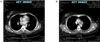
Fig. 2A, 2B
1.5 × 1.3 × 1.3 cm sized homogenous lobulated low signal intensity mass is noted on nonenhanced sagittal T1WI in pituitary stalk and suprasellar cistern, which shows heterogenous enhancement on gadolinum enhanced image.

Fig. 3A, 3B
On contrast enhanced chest CT, left breast is not visualized and conglomerated nodules with decreased attenuation
are noted in left paratracheal mediastinum and right hilum.


References
1. Weil RJ. Pituitary metastasis. Arch Neurol. 2002. 59:1962–1963.
2. Komninos J, Vlassopoulou V, Protopapa D, Korfias S, Kontogeorgos G, Sakas DE, Thalassinos NC. Tumors metastatic to the pituitary gland: case report and literature review. J Clin Endocrinol Metab. 2004. 89:574–580.
3. Kim HC, Lee HJ. A Clinical study of sellar and parasellar lesions. J Kor Neurosurg Soc. 1973. 2:37–49.
4. Jung SW, Lee KW, Kang MS, Ahn JH, Kim BS, Kim MC. Metastatic renal cell carcinoma to the hypophysis. J Kor Med Assoc. 1991. 34:671–677.
5. Shin ET, Lee EJ, Kim KR, Lee KM, Bae HD, Lee KS, Chung YS, Ahn KJ, Lim SK, Lee HC, Huh KB. A Case of pituitary metastasis from periampullary carcinoma. J Kor Endocrinol. 1993. 8:88–93.
6. Lee SH, Song YD, Kim HS, Lee YM, Yoon YS, Lim SK, Nam JH, Kwon SH, Kim KR, Lee HC, Huh KB. A Case of central diabetes insipidus caused by metastatic malignant lymphoma. J Kor Endocrinol Soc. 1997. 12:596–601.
7. Ruelle A, Palladino M, Andrioli GC. Pituitary metastases as presenting lesions of malignancy. J Neurosurg Sci. 1992. 36:51–54.
8. Morita A, Meyer FB, Laws ER Jr. Symptomatic pituitary metastases. J Neurosurg. 1998. 89:69–73.
9. Sioutos P, Yen V, Arbit E. Pituitary gland metastases. Ann Surg Oncol. 1996. 3:94–99.
10. McCormick PC, Post KD, Kandji AD, Hays AP. Metastatic carcinoma to the pituitary gland. Br J Neurosurg. 1989. 3:71–79.
11. Max MB, Deck MD, Rottenberg DA. Pituitary metastasis: incidence in cancer patients and clinical differentiation from pituitary adenoma. Neurology. 1981. 31:998–1002.
12. Houck WA, Olson KB, Horton J. Clinical features of tumor metastasis to the pituitary. Cancer. 1970. 26:656–659.
13. Teears RJ, Silverman EM. Clinicopathologic review of 88 cases of carcinoma etastatic to the putuitary gland. Cancer. 1975. 36:216–220.
14. Branch CL Jr, Laws ER Jr. Metastatic tumors of the sella turcica masquerading as primary pituitary tumors. J Clin Endocrinol Metab. 1987. 65:469–474.
15. Delattre JY, Castelain C, Davila L, Schadeck B, Poisson M. Metastasis to the pituitary stalk in a case of breast cancer. Rev Neurol (Paris). 1990. 146:455–456.
16. Chiang MF, Brock M, Patt S. Pituitary metastases. Neurochirurgia (Stuttg). 1990. 33:127–131.
17. Leramo OB, Booth JD, Zinman B, Bergeron C, Sima AA. Morley TPL Hyperprolactinemia, hypopituitarism, and chiasmal compression due to carcinoma metastatic to the pituitary. Neurosurgery. 1981. 8:477–480.
18. Schubiger O, Haller D. Metastases to the pituitary-hypothalamic axis. An MR study of 7 symptomatic patients. Neuroradiology. 1992. 34:131–134.
19. Freda PU, Post KD. Differential diagnosis of sellar masses. Endocrinol Metab Clin North Am. 1999. 28:99–100.
20. Chaudhuri R, Twelves C, Cox TC, Bingham JB. MRI in diabetes insipidus due to metastatic breast carcinoma. Clin Radiol. 1992. 46:184–188.
21. Moses AM, Clayton B, Hochhauser L. Use of T1-weighted MR imaging to differentiate between primary polydipsia and central diabetes insipidus. Am J Neuroradiol. 1992. 13:1273–1277.
22. Nelson PB, Robinson AG, Martinez AJ. Metastatic tumor of the pituitary gland. Neurosurgery. 1987. 21:941–944.
23. Yap HY, Tashima CK, Blumenschein GR, Eckles N. Diabetes insipidus and breast cancer. Arch Intern Med. 1979. 139:1009–1011.
 XML Download