

 PDF
PDF ePub
ePub Citation
 Print
Print


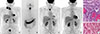
A 61-year-old man underwent 18-fluoro-2-deoxyglucose positron emission tomography/computed tomography (18F-FDG-PET/CT) staging for biopsy-proven thyroid carcinoma with right neck nodal metastases (Fig. 1A). Total thyroidectomy with central and right neck lymph node dissection was performed. Final histological diagnosis was "columnar-cell" (CC) variant of papillary thyroid cancer (PTC) and multiple nodal metastases (Fig. 1E). One month later, adjuvant radioiodine treatment (5,550 MBq) was performed, and the post-therapeutic whole-body scan showed only minimal uptake in the thyroid bed, consistent with remnant thyroid tissue (Fig 1B). At patient discharge, serum thyroglobulin was 137 ng/dL.
Three months later, a newly growing lymphadenopathy in the anterior median neck and hard subcutaneous nodules in the left cervical region were detected by a physical examination and ultrasound. They were confirmed as thyroid cancer metastases by fine-needle aspiration biopsy. For restaging purposes, the patient underwent a second 18F-FDG-PET/CT examination, which identified a suspicious lymphadenopathy in the right supraclavicular fossa (Fig. 1C, arrow) in addition to the known lesions. Serum thyroglobulin was 65 ng/dL at the time. The patient underwent revision surgery of the central and right neck lymph node compartments, in addition to left cervical nodal dissection and surgical removal of the subcutaneous nodules. Histology confirmed metastases of CC PTC in the left cervical soft-tissues (Fig. 1F) and in seven of the total 22 nodes that were removed. Based on the rapid and extended recurrence and the tumor dedifferentiation evidenced by the PET/CT findings, external-beam radiation therapy was recommended as an adjuvant treatment. A total of 44 Gy was delivered to the neck by intensity-modulated radiation therapy. Six months later, serum thyroglobulin dropped to 0.1 ng/dL and 1 year later, a follow-up 18F-FDG-PET/CT scan showed no signs of disease recurrence (Fig. 1D). CC is a rare histological subtype of PTC, usually associated with more aggressive behavior and a worse overall outcome than the classical variant [1]. In such instances, strict postoperative monitoring is recommended as the early detection and treatment of recurrence may significantly improve survival [2]. Several studies suggested that 18F-FDG-PET/CT may be a potentially valuable technique in the staging and surveillance of patients with aggressive subtypes of differentiated thyroid cancer such as the CC variant, although there is no definite consensus regarding its indications in routine clinical practice [3]. This is the first reported case of CC PTC evaluated by 18F-FDG-PET/CT, which was a useful tool in all phases of management for this patient.
 XML Download