

 PDF
PDF ePub
ePub Citation
 Print
Print


High populations of senescent fibroblasts are found in chronic ulcers as a result of oxidative stress;1 these senescent fibroblasts prevent ulcers from healing and are considered as important therapeutic targets in the treatment of chronic ulcers.2 Chronic ulcer healing can be stimulated by the administration of platelet-derived growth factor (PDGF), as PDGF restores transforming growth factor (TGF)-β signalling.345 High levels of PDGF can be obtained from the secretory factors of autologous platelets;6 clinical reports have shown that platelet gel can reduce the size of chronic venous leg ulcers and stimulate their re-epithelisation, and similar results were observed when chronic ulcers were injected with platelet-rich plasma.78
Significant populations of premature senescent fibroblasts are also found in the dermis of photoaged skin as the result of oxidative stress from chronic exposure to ultraviolet (UV) radiation, which can induce nuclear changes in human dermal fibroblasts (HDFs) by shortening telomeres and stimulating premature cellular ageing.910 In photoaged skin, collagen bundles undergo structural changes11 due to the suppression of both dermal collagen synthesis and HDF proliferation, due in turn to inhibited TGF-β1 receptor gene expression.1213 UV radiation can also stimulate collagen degradation by inducing matrix metalloproteinase-1 (MMP-1) gene expression while active collagen bundles become more sensitive to MMP-1 activity.1415
High concentrations of platelets can be obtained via platelet-rich fibrin (PRF).16 Based on the beneficial effects of platelets in the healing of chronic ulcers, we assumed that PRF could also restore the cellular functions of chronically ultraviolet-A (UVA)-irradiated HDFs in terms of cellular proliferation, collagen deposition, and cellular migration. We suggest that PRF can be developed to become an autologous material for treating photoaged skin.
To test whether PRF could restore cellular functions after chronic UVA irradiation, HDFs from the foreskins of six voluntary young human subjects (11–13 years old) were cultured in high-glucose Dulbecco's modified Eagle's medium (DMEM, Gibco, New York, NY, USA) supplemented with 10% foetal bovine serum (Gibco, New York, NY, USA), 1% penicillin-streptomycin (Gibco, New York, NY, USA), and 1% fungizone (Gibco, New York, NY, USA). These young skins were chosen as they were considered free from UV exposure and did not suffer from aging processes. The young skins were cultured until the fourth passage and divided into UVA-irradiated and non-irradiated groups. For the UVA-irradiated group, three replicates of 2×10³ HDFs from each young skin, in different 96-well plates, were exposed to 12 fluorescent lamps (L100W 79-R L UVA EVERSUN OSRAM, Munich, Germany) and treated with 3.33 J cm-2 for each irradiation treatment every 72 hours; the total dose approached 10 J cm-2. After UVA irradiation, the UVA-irradiated groups were treated with various concentrations of PRF lysate (PRF-L) diluted in DMEM supplemented with 1% foetal bovine serum. For this purpose, PRF was isolated from peripheral blood of a volunteer by a standard protocol,16 and PRF-L was isolated via incubation of PRF at 4℃ for 24 hours according to the method of He, et al.,17 2004. Meanwhile, the non-irradiated group HDFs were cultured in DMEM supplemented with 10% foetal bovine serum.
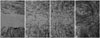
The proliferation index was calculated as the number of HDFs at the end of the experiment divided by the number of HDFs at the beginning of the experiment. Due to our trypsinized cellular harvesting procedure, it was possible that we did not collect all of the HDFs from the wells; therefore, we calculated the number of HDFs at the end of the experiment using the linear curve between optical density (OD) of the MTT [3-(4,5-dimethylthiazol-2-yl)-2,5-diphenyltetrazolium bromide, Bio Basic, Ontario, Canada] assay and the number of HDFs counted by an automated cell counter (Scepter™ Sensors, 60 um, Mexico City, Mexico). Each HDF source had its own standard linear curve. For this purpose, the duration of each MTT-assay was set at 4 hours for all experiments. The collagen deposition assay was based on the insoluble collagen of the Sirius Red assay, according to the method of Taskiran, et al.18 The levels of collagen deposition are presented as OD. Using 24-wellplates, we performed cellular migration assays as follows. After UVA irradiation, all of the bottom wells of the cultures were scratched with the blunt tip of a 32 G sterile needle through the centre of the well bottom. After incubation for 3×72 hours, the cells were fixed with cold methanol, stained with Meyer haematoxylin, and photographed (Fig. 1). Fibroblast migration rates were calculated based on the image reading method introduced by Yarrow, et al.19 Migration rates are presented in percentages. The MTT, collagen deposition, and cellular migration assays were performed concurrently.
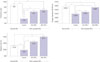
Our experiments showed that, compared to normal HDFs, UVA-irradiated HDFs showed significantly decreased (p<0.05) proliferation indexes (8.61±0.64 vs. 3.33±0.89, respectively), collagen deposition (0.584±0.031 OD vs. 0.393±0.028 OD, respectively), and cellular migration (31.69±2.54% vs. 18.84±1.74%, respectively). UVA-irradiated HDFs cultured in 25% and 50% PRF-L showed significantly higher proliferation indices, collagen deposition, and cellular migration rates than cells in diluent medium, although their proliferation indices remained less than those of normal fibroblasts (Fig. 2).
Repeated UVA irradiation can induce cellular senescence and stimulate MMP-1 mRNA expression in HDFs.10 Similar events were also found in our experiments, specifically that UVA irradiation of HDFs can significantly inhibit (p<0.05) the cellular proliferation index, collagen deposition, and migration, similar to fibroblasts in prematurely aged skin, possibly due to either cellular senescence or UVA-induced decreasing global cellular function. Unfortunately, due to our limitations, evidence of cellular senescence, such as senescence-associated galactosidase staining, was not collected in this experiment.
Incubation of platelet concentrate at 4℃ for 24 hours produces high concentrations of PDGF-BB, epidermal growth factor, and TGF-β1;20 similar findings have been found with PRF-L.17 Those growth factors effectively stimulate both collagen and hyaluronic acid production by dermal fibroblasts and likely help treat skin aging.21 Our findings demonstrated that 50% PRF-L can significantly improve the proliferation index, collagen deposition, and migration rates of chronically UVA-irradiated HDFs (p<0.05) (Fig. 2), compared to diluent medium. Fig. 2 shows that the treated groups had the same collagen deposition as normal HDFs and significantly better migration (p<0.05); 50% PRF-L failed to achieve normal HDF proliferation indices (p<0.05), and 25% PRF-L could only ameliorate the migration rates of chronically UVA-irradiated HDFs.
As mentioned above, decreased TGF-β1 receptor gene expression among the fibroblasts in photoaged skin leads to impaired TGF-β1 signalling and impaired cellular proliferation, collagen synthesis, and migration. We assume that the repaired proliferation indexes, collagen deposition, and migration of chronically UVA-irradiated HDFs were due to amelioration of TGF-β1 signalling, based on the results of previous experiments on dermal fibroblasts and vascular smooth muscle cells.45 On the surface of the dermal fibroblast membrane, the receptors for PDGF-BB and TGF-β1 both physically interact and ameliorate each other's signalling and stability.22 Another factor responsible for the lack of collagen content in prematurely aged skin is MMP-1 gene expression for collagen type-1 degradation; a previous study showed that platelets can release tissue inhibitors of MMP.23 Therefore, we also assume that the improved collagen deposition of UVA-irradiated HDFs is caused by the inhibition of MMP-1 activity by tissue inhibitors of MMP in PRF-L. We do not have evidence to support these assumptions in our experiments; therefore, further experiments are necessary.
As PDGF has a molecular weight of 35 kD and therefore cannot be absorbed through the skin surface, direct application of autologous PRF-L on intact skin is useless. Learning from the success of mesotherapy skin rejuvenation with hyaluronic acid, intradermal injection with PRF-L using a mesogun can be attempted in patients with aged skin.24 Another clinical experiment is the cutaneous application of PRF-L after the application of glycolic acid to enhance skin penetration, such as in the clinical study of a eutecticmixture of local anaesthetic.25
In conclusion, PRF is a good candidate material for treating prematurely aged skin; however, more clinical trials are needed to establish an appropriate application method of this material.
 XML Download