

 PDF
PDF ePub
ePub Citation
 Print
Print


INTRODUCTION
Recent epidemiologic studies increased our understanding on the role of inflammation played in atherosclerosis and cardiovascular disease (CVD)1-4 and considerable amount of interest has also been focused on the positive relationship between inflammation and metabolic syndrome (MS).5,6 Inflammation is known to be associated with the pathogenesis of MS itself as well as coronary heart disease,7,8 and proinflammatory cytokines play important roles in the relation between MS and CVD. Recent studies have also demonstrated that high serum C-reactive protein concentrations are associated with MS.9,10
White blood cell (WBC) count is an objective marker of acute infection, tissue damage, and other inflammatory conditions. An increase in the WBC count is closely related with advanced atherosclerosis and the incidence and mortality rate of CVD.11 In cross-sectional studies, WBC counts are correlated with isolated variables of MS and clearly increased with numbers of MS.5,6,12,13 In addition, Oda and Kawai14 reported that the prevalence of MS and diabetes increases through the quartiles of WBC in Japanese men and women. Although several epidemiological studies have already noted a relationship between WBC counts and MS, most of these studies are cross-sectional studies. Few studies have been performed to evaluate the relationship between baseline WBC count and future risk for developing MS.15,16
Therefore, we investigated whether the baseline plasma levels of the WBC count could impact the future risk for MS in apparently healthy Korean adults.
MATERIALS AND METHODS
Subjects
We conducted a retrospective study of 1476 participants who underwent annual health examinations at Kangbuk Samsung Hospital in both 2002 and 2005. The subjects were inhabitants of either Seoul (capital of South Korea) or Gyeonggi province (satellite town). We excluded those subjects who already met the MS criteria at baseline (n=217) and those subjects with other missing data for components of metabolic syndrome. The subjects whose WBC counts beyond 10400/µL were also excluded from the study. The subjects with documented acute infection and malignancy were also excluded. Finally, we analyzed 1135 subjects (781 males, 354 females). Informed consent requirement for this study was exempted by the institutional review board because researchers accessed only the database for analysis, and personal information was not accessed. This study was approved by the institutional review board at Kangbuk Samsung Hospital.
Measurements
The participants underwent routine physical examinations that included the measurement of height, weight, blood pressure, and overnight fasting blood sampling. The standing height was measured without shoes to the nearest 0.1 cm. Weight was determined without shoes on a light cloth. Blood pressure was measured by using a standard mercury sphygmomanometer after being seated for at least 5 min. Blood was sampled after 12 hours of fasting, and the venous blood glucose, insulin, total cholesterol (TC), triglyceride (TG) and high- and low-density lipoprotein cholesterol (HDL-C and LDL-C, respectively) were measured. Plasma glucose was measured in duplicate by the hexokinase method using an autoanalyzer (Hitachi, Tokyo, Japan), which had an interassay coefficient of variation of 1.6%. An enzymatic calorimetric test was used to measure the TC and TG levels. The selective inhibition method was used to measure the level of HDL-C and a homogeneous enzymatic calorimetric test was used to measure the level of LDL-C. The fasting serum insulin level was measured by immunoradiometric assay (RIABEAD II, Abbott, Tokyo, Japan) having an intra-assay coefficient of variance of 1.2% to 1.9% and an inter-assay coefficient of variance of 1.4% to 3.3%. Insulin resistance status was calculated by using the homeostatic model assessment-insulin resistance (HOMA-IR). HOMA-IR=[fasting insulin (µIU/mL)×fasting blood glucose (mmol/L)]/22.5. The total WBC count and differential count were assessed by an autoanalyzer (ADVIA 129, Bayer, Germany). The presence of MS was defined by using a modified version of the ATP III criteria.17 Briefly, four of the five MS components were defined using the following ATP III categorizations: 1) high blood pressure (BP), ≥130/85 mm Hg or patients using anti-hypertensive agents; 2) Hypertriglyceridemia, ≥150 mg/dL (1.693 mmol/L); 3) Low HDL-C, <40 mg/dL (1.034 mmol/L) in men and <50 mg/dL (1.293 mmol/L) in women; 4) High fasting glucose, ≥100 mg/dL (5.6 mmol/L). The fifth component was defined based on body mass index (BMI) because waist circumference was not available in all subjects. Central obesity was diagnosed at a BMI (≥25 kg/m2). The subjects with three or more of the following criteria were defined as having MS. The smoking status of subjects was classified as non-smoker or smoker (former or current).
Statistical analysis
Statistical analyses were performed by SPSS version 11.0. All values were presented as mean±SD. Comparisons of WBC counts according to two groups who have MS or not were performed by independent sample t-test. The chi-square test was used for categorical variables. WBC counts were divided into quartiles and logistic regression analysis was used to evaluate the risk for each category of WBC count for the association with MS. For each risk, a 95% confidence interval was determined. Also, quartile-specific risk estimates were obtained through logistic regression after adjusting for age, gender, smoking, BMI, HOMA-IR and other components of MS. Differences in the prevalence of MS according to the quartiles of WBC count were determined by chi-square test. p values of <0.05 were considered statistically significant.
RESULTS
Clinical characteristics
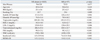
Comparisons of the baseline characteristics of the subjects who were subsequently developed MS and did not develop MS are shown in Table 1. The mean age of the subjects were 49 years. Subjects who were subsequently diagnosed with MS in 2005 had a higher baseline BMI, systolic BP, diastolic BP, triglyceride, T-cholesterol, fasting glucose, fasting insulin and HOMA-IR. The subjects who did not develop MS in 2005 had a higher baseline HDL-C. The mean baseline levels of the WBC were significantly higher for the MS group (p<0.001). The overall present incident MS in 2005 was 8.9%.
Clinical characteristics according to the quartiles of WBC count
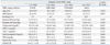
We classified subjects according to the quartiles of WBC count and their clinical characteristics were compared (Table 2). BMI, triglyceride, LDL-C, total cholesterol, fasting glucose, fasting insulin and HOMA-IR increased significantly from the 1st quartile to the 4th quartile of WBC count. HDL-C decreased significantly from the 1st quartile to the 4th quartile of WBC count.
Risk of developing MS according to the quartiles of baseline WBC levels
The relative risks for MS according to the quartile groups of WBC count are shown in Table 3. In unadjusted models, the relative risks of incident MS in 2005 were 1.4, 3.2 and 2.7 for the WBC quartiles 2, 3, 4 when compared with first quartile (p-value for trend <0.001). After further adjustment for HOMA-IR as index of insulin resistance, the relative risks of incident MS in 2005 were 1.1, 2.5 and 2.1 for the WBC quartiles 2, 3 and 4, respectively, when compared with the first quartile (p-value for trend=0.011). Additional multivariable adjustments for components of MS i.e., baseline BMI, BP, fasting glucose, HDL-C and triglycerides, attenuated these associations, but these positive associations persisted (Table 3).
DISCUSSION
This retrospective cohort study indicates that an elevated level of WBC is associated with later risk of developing MS in apparently healthy Koreans. Several cross-sectional studies have reported the association between WBC and MS.12-14,18-20 Wang, et al.5 reported that subjects in the highest quartile of WBC counts demonstrated a threefold increase, in the odds ratio for MS compared to subjects in the lowest quartile of WBC counts. Also, in the study of Lao, et al.,19 the risk of MS increased significantly with higher total WBC, and vascular risk factors were positively associated with WBC in older Chinese, and Lohsoonthorn, et al.13 showed that, the risk of MS increased across the successive quartiles of the WBC counts among Thai women. Finally, Kim, et al.20 reported that the OR for MS was 2.64 in the highest quartile of total WBC count versus the lowest quartiles of commensurate total WBC count in a Korean population. However, all of these studies are cross-sectional studies. Only few studies have been performed to evaluate the association between baseline WBC counts and future development of MS. Odagiri, et al.15 investigated the 7-year incidence of MS in relation to quartiles of WBC in healthy workers, and found that the incidence of MS was 1.2 to 1.6 times higher in the higher quartile of the WBC compared with the lowest quartile.
Although the mechanisms involved in the linking of WBC counts with MS remain unclear, there are several postulations. Some authors proposed that insulin resistance or hyperinsulinemia, an essential component of MS, probably results from the chronic activation of the immune system and inflammation.21 Proinflammatory cytokines such as tumor necrosis factor-alpha (TNF-α) and interleukin-6 (IL-6), in addition to being produced by activated macrophages, were shown to be appreciably synthesized in adipose tissue,22,23 and TNF-α and IL-6 secreted from adipose tissue were shown to impair the action of insulin on adipocytes and muscle cells by paracrine/autocrine manner.23,24 Because WBC counts are increased by cytokines and are closely associated with both insulin resistance and hyperinsulinemia, their association could result in a subclinical inflammatory reaction and the expression of an insulin-resistant state.25 In the present study, we also showed that fasting insulin and HOMA-IR increased according to the increasing of WBC quartiles, in agreement with the findings that WBC count is associated with insulin resistance and insulin concentration.26 Nevertheless, the risks for MS, as calculated according to baseline WBC levels' quartile distribution in the present study, persisted after adjustment for HOMA-IR. Future large scale study is needed to clarify underlying pathophysiological role of WBC played in the genesis of MS.
Because our study population included subjects who frequently received health check-up, several characteristics of the present study population may be different from other populations that do not receive available medical services. We think that most of the study subjects are in economic stability. Therefore, the generalizability of our study may be limited. In the present study, the prevalence of MS in 2005 was 8.9%, which is seemingly low compared with those values reported in other ethnic groups. There is a possibility that the participant bias might have affected these results.
Studies regarding inflammation and CVD have been confounded by the fact that cigarette smoking increases numerous inflammatory markers, including leukocyte counts.27,28 However, in several large-scale studies, there is positive correlation of total WBC count with CVD that is independent of smoking.6,29 Also, we found that increased WBC level was significantly associated with later risk of developing MS, even after adjusting for smoking.
The present study has some limitations. First, the subjects were urban habitants (either Seoul or Gyeonggi province). The participants were the employees of the company, whose annual health check-up was carried out in the health promotion center or their family members and most of the subjects were likely members of upper-middle economic class. Although it could be regarded as the strength of this study, the participants of this study were a group of relatively homogeneous characters:, a follow-up cohort of annual health check-up program in a single health promotion center in Korea, and not the representative of the ethnic group. Therefore, the generalizability of our study may be limited. This could have biased the baseline characteristics of the study cohort and future development of MS.
Second, the serum high-sensitivity C-reactive protein (hsCRP), a marker of chronic low-grade inflammation, is known to be a sensitive predictor of CVD and is related to components of MS. Comparison of WBC counts and hsCRP levels is needed for more definitive conclusion on the usefulness of WBC as an independent predictor of incident MS, since our study did not include hsCRP levels. The strengths of our study would be higher if we performed this analysis.
Third, we used BMI instead of waist circumference as criteria of central obesity. This might underestimate the effect of obesity on MS. Various diagnostic criteria of MS have been proposed by different organizations over the past decade.30 The main difference concerns the measure for central obesity. Indeed, there was no agreement on the definition of abdominal obesity between the International Diabetes Federation (IDF) and the American Heart Association/National Heart, Lung, and Blood Institute.31 Alberti, et al.31 indicated that waist circumference differs in different populations and may require modification for different ethnic group. The IDF values are closer to a BMI of 25≥ kg/m2 in males. Recent studies have used BMI successfully to classify those with MS, lending support to the validity of our measure.32,33 In particular, Shiwaku, et al.34 stated that the definition of BMI 25≥ kg/m2 as obesity is suitable for the determination of the MS among Korean and Japanese.
Fourth, we didn't exclude patients who were positive for hepatitis B virus surface antigen and/or hepatitis C virus antibody and with helicobacter pylori infection, which are prevalent in Korea.
Fifth, our study lacked information on drug use, which could affect serum WBC counts including antidiabetic, antihypertensive, or statin medication. The strengths of our study would be higher if we excluded these confounding factors.
In conclusion, in the present study, increased WBC level was associated with later risk of developing MS in apparently healthy adults. Further large-scale prospective studies are warranted to clearly reveal the underlying pathophysiological role of WBC for MS and to establish better clinical utility to predict the development of MS.

 XML Download