

 PDF
PDF ePub
ePub Citation
 Print
Print


INTRODUCTION
Since the first report by Carlson, et al.,1 reexpansion pulmonary edema (REPE) has been recognized as a potentially fatal yet uncommon complication occurring after rapid expansion of the lung after closed thoracostomy in patients with hemothorax, pneumothorax or pleural effusion.2 In previous studies, the prevalence of REPE has varied between 0.9% to 57%. Recognition of REPE has become more common with the development of improved diagnostic modalities such as computerized tomography.3-5 Management of patients with pneumothorax is most commonly by closed thoracostomy. Closed thoracostomy may be performed by one of two methods. The first uses a trocar to insert a chest tube into the pleural cavity while the second uses a hemostat to enter the pleural cavity. The major potential advantages of the trocar-assisted method include its simplicity, rapid procedure time, and its ability to minimize associated tissue injury, pain, and subsequent scarring. In contrast, the hemostat-assisted method is thought to be safer though slower than trocar-assisted thoracostomy.
A number of studies have explored potential risk factors for REPE after drainage of spontaneous pneumothorax, however, the association between REPE and the method of closed thoracostomy has not yet been reported. The objective of the current study was to explore possible association between the method of closed thoracostomy and REPE after drainage of spontaneous pneumothorax.
MATERIALS AND METHODS
Study design
A prospective observational study was conducted to determine clinical characteristics and frequency of REPE, based on the method of closed thoracostomy used to treat spontaneous pneumothorax. The study was approved by the Institutional Review Board and all patients gave informed consent.
Setting and subjects
The study was conducted at a suburban emergency department that is part of an academic medical center in Korea. Patients presenting to the Wonju Christian Hospital with a spontaneous pneumothorax who were treated with either hemostat- or trocar-assisted closed thoracostomy between January 2007 and December 2008 were eligible for enrollment.
Measures and outcomes
Structured data collection was performed on study patients. All patients underwent an erect posteroanterior (PA) or supine anteroposterior (AP) radiograph of the thorax to confirm the presence of a spontaneous pneumothorax. Following closed thoracostomy, the end of the thoracostomy tube was connected to a single bottle system that was attached to a negative pressure (20 cm H2O using an Emerson post-operative suction pump model 55-JS, J. H. EMERSON Corp., Cambridge, MA, USA). The size of the pneumothorax was classified into one of the following four groups: small, medium, large and tension pneumothorax. A small pneumothorax was defined as a pneumothorax that was localized in the apex of the lung on chest radiography. A medium pneumothorax was defined as a pneumothorax that extended beyond one third of the width of a hemithorax. A large pneumothorax was defined as a pneumothorax leading to complete or nearly complete collapse of the lung parenchyma. A tension pneumothorax was defined as a pneumothorax associated with depression of diaphragm, or a shift of mediastinum and trachea away from the collapsed lung.5 PaO2, PCO2, SaO2, lactate, WBC count, troponin I and b-type natriuretic peptide were measured in all patients prior to tube thoracostomy. Subsequent radiographs and chest computed tomography (CT) scans were obtained within 8 hours of closed thoracostomy to evaluate the need for bullectomy and verify the extent of reexpansion of collapsed lung and the presence or absence of pulmonary edema of the involved lung parenchyma. The determination of the presence of REPE was made by two investigators (a board-certified emergency physician and a board-certified radiologist) who were blind to demographic and clinical information.
Estimation of pneumothorax size
The average interpleural distance (AID) was calculated in chest X-ray (CXR) as the arithmetic mean of the maximum apical interpleural distance (A) and two measurements of interpleural distances (B and C) at the midpoints of the upper and lower halves of the lung.6
In erect PA radiographs, the percentage of pneumothorax volume was obtained from the following formula: Percentage pneumothorax in erect PA radiograph=4.2+[4.7×(A+B+C)].7
Due to severe dyspnea and pain, some patients were not able to maintain an erect position during chest radiography, and only supine radiographs were performed in these patients. The above formula for calculating the size of pneumothorax could not be applicable with chest radiographs obtained in the supine position, in children (0-12 years old), or in patients with hydropneumothoraces.8 When supine radiographs were used, the percentage of pneumothorax volume was calculated using the following formula: percentage pneumothorax on supine AP radiograph=8.73+10.03×AID.9
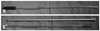
Closed thoracostomy techniques (Fig. 1)
During the first year of the study (2007), all study patients underwent hemostat-assisted thoracostomy while patients presented during the second year (2008) underwent trocar-assisted thoracostomy.
The hemostat-assisted technique was performed using the following sequence:
A 2 cm-length skin incision was made at the rib level below the pleural puncture site.
Intercostal muscle dissection was performed using a straight hemostat.
Penetration of the parietal pleura was performed using a hemostat, and a thoracostomy tube (P.V.C Thoracic Catheter, 28 Fr., Sewoon Medical Co., Ltd., Cheonan, Korea) was inserted into the pleural cavity.
The trocar-assisted technique was performed using the following sequence:
Following closed thoracostomy, the tube was connected to a single bottle system that was attached to a negative pressure (20 cmH2O using an Emerson post-operative suction pump, model 55-JS, J. H. Emerson Corp., Cambridge, MA, USA).
Data analysis
Data were analyzed using SPSS 12.0 for Windows statistical software package (SPSS, Chicago, IL, USA). Continuous data are presented as means with standard deviations and compared with the independent sample t-test or Mann-Whitney U test if appropriate. Nominal data are presented as the percent frequency of occurrence and compared with a χ2 or Fischer's exact test when appropriate. The association between potential predictor variables and the occurrence of REPE was determined using logistic regression analysis. The level of significance was preset at a p value of 0.05.
RESULTS
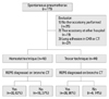
From January 2007 through December 2008, we prospectively evaluated 173 patients who were diagnosed with a spontaneous pneumothorax. Eighty-one patients were excluded since they were treated by 100% oxygen inhalation alone, were transferred from another hospital after tube thoracostomy, or had extensive adhesions on radiography (Fig. 2).
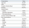
The study sample included 83 males with a mean age of 25±12 years. The mean time interval from symptom onset to ED visit was 2.2±4.1 days. Of all pneumothoraces, 20 were small, 20 were medium, 49 were large, and 21 were tension pneumothoraces.
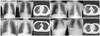
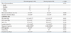
Of all pneumothoraces, 41 (45%) involved the right lung and hemostat-assited thoracostomy was performed in 48 patients (52%). Based on subsequent CT imaging of the chest, REPE developed in 68 patients (74%) (Fig. 3) (Table 1). The frequency of REPE was higher in patients undergoing trocar-assisted thoracostomy (38 patients, 86%) than those undergoing hemostat-assisted thoracostomy (30 patients, 63%; p=0.009). Age, gender, time interval from symptom onset to ED visit, location of pneumothorax, size of pneumothorax, time interval from thoracostomy to CXR evaluation, and time interval from thoracostomy to CT evaluation were not different between the two groups. There were also no between-group differences in PaO2, PCO2, SaO2, and lactate.
All of the study patients were admitted to the hospital and there were no subsequent major complications or deaths due to the development of REPE. Bullectomy was performed in cap28 patients undergoing hemostat-assisted thoracostomy (58.3%) and in 28 patients undergoing trocar-assisted thoracostomy (63.4%). Mean length of hospital stay was 6.5±3.2 days after hemostat-assisted thoracostomy and 6.6±3.5 days after trocar-assisted thoracostomy (p=0.930) (Table 2).
We performed logistic regression analysis to determine the association between potential contributing factors and the development of REPE. The only factor significantly associated with REPE was trocar-assisted thoracostomy [odds ratio (OR) 5.7; 95% confidence interval (CI) 1.5 to 21.4; p=0.009]. The size of pneumothorax had little impact on the development of REPE (OR=1.1, 95% CI 1.0 to -1.1; p<0.001). Age, gender, and time interval from symptom onset to ED visit had no impact on the development of REPE (Table 3).
DISCUSSION
Our results demonstrate that while the frequency of REPE was increased when the trocar technique was used for closed thoracostomy in patients with spontaneous pneumothorax, it did not affect the ultimate outcome of the patients in this study.
The different rates of REPE between the two methods of closed thoracostomy might be due to differences in the rate of re-expansion of the collapsed lung. In an animal study, the investigators hypothesized that rapid decompression and re-expansion of the collapsed lung could result in capillary vascular injury and ipsilateral pulmonary edema while more gradual decompression might prevent pulmonary edema.10,11 When the hemostat is used to decompress a pneumothorax, decompression of the collapsed lung may be more gradual. The first phase of decompression occurs when the intercostal muscle and the parietal pleura are dissected. A second decompression phase occurs when the thoracic cavity is entered with a finger. The last phase of decompression occurs when the thoracostomy tube is inserted into the pleural cavity using a hemostat. In contrast, the trocar technique is performed in one step with only a minimal skin incision and direct puncture and insertion of the thoracostomy tube through the parietal pleura. As a result, intrathoracic pressure may decrease more rapidly with the trocar technique and thus could contribute to rapid reexpansion of the collapsed lung, capillary injury and more frequent development of REPE.
Several investigators have hypothesized that REPE is the result of pulmonary vascular injury and an increase in capillary permeability. Carlson, et al.1 earlier reported that pulmonary capillary permeability increased when pulmonary circulation was normalized after hypoxic vascular injury during lung collapse. An increase in nitric oxide or xanthine oxidase after reexpansion of the collapsed lung was thought to result in an increase in pulmonary capillary permeability.12,13 The change in surface tension after long-standing collapse of lung has also been suggested to result in decreased surfactant production and pulmonary edema.14
Several risk factors for REPE have been proposed, including chronicity of collapse, larger size of pneumothorax, younger age, and underlying diseases of patients.4 To the best of our knowledge, however, no prior study has investigated the association between the method of thoracostomy and the development of REPE. Since we excluded patients with underlying lung adhesions, most of our cases had no underlying pulmonary diseases, and this may explain why we did not identify any such risk factor for REPE.
The detection rate of REPE was higher in our study than other earlier studies, possibly due to the application of negative pressure after thoracostomy and the use of CT imaging to detect REPE.15 CT imaging of the chest was used in this study, because of its higher accuracy and sensitivity in diagnosing REPE. As a result, even small and asymptomatic cases of REPE were identified. In contrast, earlier studies have used CXR for the diagnosis of REPE, therefore, the incidence of REPE in our study was higher than previous studies, and it may be a more accurate reflection of true incidence of REPE after closed thoracostomy.
It is well known that most patients with REPE have no significant complications and do not require specific treatment modalities such as assisted ventilation or ICU care.2 All of the patients with REPE in our study also recovered after conservative management alone without any complications. However, REPE-related mortality due to acute respiratory failure has been reported in several studies, and physicians should continue to be concerned with the clinical course of patients with REPE.8
The major limitation of our study is the fact that most of the clinical variables were limited to the ED and there was little information on the clinical course of pulmonary edema after hospital admission. Therefore, we could not evaluate whether the development of REPE was related to deterioration of clinical symptoms such as dyspnea, tachypnea or palpitation during hospital stay.
Second, Emerson pump with a negative pressure of 20 cmH2O was applied to all enrolled patients. Rapid negative pressure application in long term lung collapse might be related to increased incidence of REPE.3,11 Thus, it is likely that the use of Emerson pump with negative pressure may affect to the incidence of REPE. However, REPE can occur in patients without any negative pressure suction.16 Third, we used chest CT for diagnosing REPE instead of plain chest X-ray. It could have overestimated the incidence of REPE. However, chest CT is superior to chest X-ray for diagnosing the REPE, we had used the most sensitive imaging tool.
Forth, we used single size thoracostomy tube in both groups regardless of pneumothorax size. Various thoracostomy techniques by pneumothorax size are introduced in recent studies.17-19 However, we used single size tube in order to exclude decompression pressure variation by diameter of pleural opening. Nevertheless, the opening of the pleura might be bigger in hemostat technique because the opening of the pleura was made manually prior to insertion of thoracostomy tube. Thus, decompression pressure could be different in two groups and it could affect the frequency of REPE. Fifth, patient allocation was not randomized. This may have led to the introduction of additional biases. Finally, while measuring the size of the pneumothorax with CT would have been more accurate than with CXR based formula,20,21 most patients had CT imaging after thoracostomy because of cost and patient convenience.
In conclusion, the incidence of REPE in ED patients with spontaneous pneumothorax was higher after trocar-assisted thoracostomy, however, ultimate clinical outcome was similar to hemostat-assisted thoracostomy. When using the trocar technique for the treatment of spontaneous pneumothorax, intermittent clamping of thoracostomy tube to slow reexpansion of the collapsed lung should be considered.

 XML Download