

 PDF
PDF ePub
ePub Citation
 Print
Print


INTRODUCTION
Sarcomas are a heterogeneous group of rare tumors that arise predominantly from the embryonic mesoderm and account for about 1% of all malignant tumors.1 Soft tissue sarcoma of the genitourinary tract are rare, accounting for only 2.1% of soft tissue sarcomas and 1% to 2% of all malignant genitourinary tumors.2,3 Due to their rarity, there are very few studies on genitourinary sarcomas in the literature,4,5 and little is known about their natural history and prognosis. In the present study, we analyzed the clinicopathological characteristics and prognosis of 18 cases of localized resectable genitourinary sarcomas in adults that were collected in a single institution. To the best of our knowledge, this is the first study of genitourinary sarcomas in adults in an Asian population.
MATERIALS AND METHODS
Patients
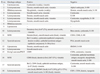
Approval to perform this study was obtained from the institutional review board. Between September, 1996 and November, 2008, consecutive adult patients who were treated for primary genitourinary sarcomas at our hospital were identified through a database search. Their medical records were thoroughly reviewed for pertinent information regarding the presentation, tumor characteristics, therapy, and outcome. Patients with distant metastases were excluded from the analysis. The patient evaluation included a complete medical history, physical examination, laboratory investigations, and radiologic imaging data (chest X-ray, computerized tomography and/or magnetic resonance imaging, and bone scans). Preoperative staging revealed that none of the patients had metastatic disease at presentation. Our series only included sarcomas of the genitourinary tract such as kidney, ureter, bladder, prostate, urethra, and paratesticular region. Retroperitoneal sarcomas involving the genitourinary organs were excluded from the analysis. The genitourinary sarcomas were characterized on the basis of positive and negative staining for specific immunohistochemical markers (Table 1). All cases were classified according to the French Federation of Cancer Center System Grading Scheme for Adult Sarcoma. In the French System, three parameters are scored individually, namely, tumor differentiation, mitotic activity, and the degree of tumor necrosis. These individual scores are summed up to yield a final score (I, II, or III) that indicates the grade of the sarcoma.6
Statistical analysis
The date of surgery served as the start of observation, and recurrence-free survival and disease-specific survival were the study end-points. Recurrence was defined as recurrent disease at a local or distant site. Death caused by disease was the only end-points for disease-specific survival. The actuarial probability of these end-points was modeled by the Kaplan-Meier method. Univariate analysis of variables associated with survival end points was performed by using the log-rank test. The variables that were analyzed were patient age, gender, body mass index, American Society of Anesthesiologists (ASA) score, primary organ, tumor histology, size, necrosis, Fédération Nationale des Centres de Lutte Contre le Cancer (FNCLCC) grade, and surgical margin positivity. With regard to the continuous variables, the patients were stratified into two groups: for age, two groups were ≤50 years and >50 years; for body mass index, they were ≤23.0 kg/m2 and >23.0 kg/m2; for ASA score, they were 1 or >1; for tumor size, they were ≤5 cm and >5 cm; and for FNCLCC grade, they were 1 or >1. Tumor location was categorized as kidney and others, and histologic subtype was categorized as leiomyosarcoma and others. Due to the small sample size, multivariate models were not appropriate.7 For all statistical analyses, p<0.05 was considered significant. All statistical tests were performed by the Scientific Package for Social Sciences software.
RESULTS
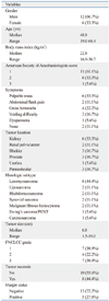
The characteristics of the 18 patients are summarized in Table 2. There were 12 males and six females. The mean and median ages at presentation were 47.8 and 48.8 years, respectively. The most common presenting symptom was a palpable mass (33.3%), and the most common site was kidney (six cases, 33.3%), followed by bladder (three, 16.7%), prostate (three, 16.7%), paratesticular region (three, 16.7%), renal pelvis/ureter (two, 11.1%) and urethra (one, 5.6%). The most common histological subtype was leiomyosarcoma (eight patients, 44.4%), followed by liposarcoma, rhabdomyosarcoma, synovial sarcoma, malignant fibrous histiocytoma (MFH) [two patients (11.1%) each], and others (two patients, 11.1%). Complete resection with negative surgical margins was achieved in 13 patients (72.2%)
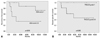
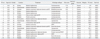
The outcome of the patients with genitourinary sarcoma is shown in Table 3. Of the 18 patients, none underwent neoadjuvant treatment. The median follow-up period for the 18 patients was 49.9 months (mean 62.9; range 6.4 to 147.6). Of all patients, six patients (33.3%) developed recurrent disease at a median time to surgery of 17.9 months (mean 28.6; range 4.4 to 89.0). The recurrence-free survival rate at 1, 3, and 5 years was 81.6%, 66.5%, and 66.5%, respectively. On univariate analysis, recurrence-free survival significantly associated only with ASA score (Fig. 1A). At a median follow-up period of 4.0 years, ten patients (55.6%) had no evidence of disease, two (11.1%) had disease, five (27.8%) had died of the disease, and one (5.6%) had died of other cause. The disease-specific survival rate at 1, 3, and 5 years was 88.9%, 76.2%, and 67.7%, respectively. On univariate analysis, disease-specific survival was significantly associated only with FNCLCC grade. Fig. 1B depicts the disease-specific survival curve of the 18 patients according to FNCLCC grade.
DISCUSSION
Sarcomas of the kidney
Two of our patients had leiomyosarcomas of the kidney. Leiomyosarcoma accounts for 50-60% of all renal sarcomas, therefore, it is the most common histologic subtype.8 Renal leiomyosarcoma has a very poor prognosis, with most patients dying within two years,9,10 but radical surgery seems to offer the best chance of cure. The role of adjuvant chemotherapy and/or radiotherapy remains debatable due to the paucity of data on the treatment of this rare renal neoplasms. While local recurrence is common, low-grade leiomyosarcoma tends to pursue a more indolent course. Of our cases, two patients had a low-grade leiomyosarcomas and no evidence of local recurrence or distant metastasis was noted during 57.2 and 147.6 months of follow-up.
One of our patients had primary renal synovial sarcoma, which is an extremely rare neoplasm that was discovered only relatively recently. Regardless of the treatment, the prognosis of renal synovial sarcoma remains poor. Indded, our case expired 6.4 months after radical nephrectomy.
Our another patient had renal carcinosarcomam which tended to be large and bulky due to its rapid growth and wide infiltration. It is known to progress rapidly and is associated with a poor prognosis. Kuroda, et al.11 have reported the case pf a patient with renal carcinosarcoma who died of systemic metastases 5 months after radical nephrectomy. The tumor of our patient also recurred 4.4 months after radical nephrectomy; the patient remains alive with disease 8.9 months after surgery.
The remaining two patients of our series with kidney sarcomas had Ewing's sarcoma/primitive neuroectodermal tumor (PNET) and MFH, respectively. Both are also characterized by an aggressive clinical course and a poor prognosis. Indeed, both of our patients had a poor outcome: one was dead from the disease and the other is alive with the disease.
Rhabdomyosarcoma of the renal pelvis
One of our patients had rhabdomyosarcoma of the renal pelvis. This is a very rare renal pelvis neoplasm, especially in adults, with only two cases of renal pelvis rhabdomyosarcoma in adults being reported to date.12,13 With regard to pathological subclassification, these tumors were of embyronal and pleomorphic type, respectively. Our case was found to have pleomorphic rhabdomyosarcoma. He was subjected to radical nephrectomy, after which he received 18 cycles of post-operative chemotherapy with the vincristine, adriamycin, cyclophosphamide (VAC)/ifostamide and etoposide (IE) regimen. At the 9-year follow-up, he is still alive without disease recurrence.
Leiomyosarcoma of the ureter
One of our patients had leiomyosarcoma of the ureter. A recent literature review found less than 20 cases of primary leiomyosarcoma of the ureter.14 Generally, as indicated by the short survival of the reported cases, the prognosis of this disease is unfavorable. Our patient was treated surgically by ureterectomy and the cutting margin of the ureter was negative. Adjuvant radiation therapy was given after surgery and the patient remains free of the disease 108 months after surgery. Although adjuvant chemotherapy has not yet been proven to be effective and remains to be tested, wide local excision and radiation therapy could be the treatment of choice for leiomyosarcoma of the ureter.14
Leiomyosarcoma of the urinary bladder
Our series contained three cases of leiomyosarcoma of the urinary bladder. To date, over 200 cases of bladder sarcomas have been reported. Of these, 50% were leiomyosarcomas, 20% were rhabdomyosarcomas, and the remainder consisted of other histologies, namely carcinosarcomas, angiosarcomas and osteosarcomas.15 Spiess, et al.16 did not detect any differences in disease-specific and overall survival durations between patients with bladder leiomyosarcoma and other sarcoma subtypes. Analysis of M.D. Anderson Cancer Center data showed that bladder sarcoma is associated with a 5-year disease-specific survival rate of 59.0-62.0%.16,17 Thus, bladder leiomyosarcoma is thought to have a poor prognosis. However, Rosso, et al.18 reviewed their experiences at the Memorial Sloan-Kettering Cancer Center, stating that some patients with bladder leiomyosarcoma are able to achieve long-term survival. They also reported a remarkable 5-year disease-specific survival rate of 84.0%. In the present study, all three patients with bladder leiomyosarcoma underwent partial cystectomy as the only mode of treatment. One patient continues to do well without evidence of disease recurrence, but two patients died of the disease 29.6 months and 27.9 months after surgery, respectively. Interestingly, the patient with no evidence of disease had positive margins after surgery, and adjuvant treatment was not advocated. However, as shown for other soft tissue sarcomas, patients with positive surgical margins can be candidates for adjuvant radiotherapy.19 Thus, our experiences suggest that leiomyosarcoma of the bladder should be treated by radical cystectomy if surgical resection is possible, while partial cystectomy should be considered as a palliative treatment, even when free margins are achieved.
Sarcomas of the prostate
Three of our patients had sarcomas of the prostate. The rare occurrence of prostate sarcomas in adults means that the survival rates associated with this disease can be inferred only from anecdotal experiences; its rarity has also limited the development of effective treatment strategies. Surgery has been the mainstay of treatment and usually involves cystoprostatectomy or total pelvic exenteration. Historically, the long-term survival rates for adult patients with prostate sarcoma are poor. However, Sexton, et al.20 suggest that a combined multimodality approach, where surgery is the mainstay of treatment and usually involves cystoprostatectomy, may result in improved survival rates. The 1-, 3- and 5-year acturarial survival rates of their 21 patients were 81%, 43% and 38%, respectively. In our series, one patient, who had rhabdomyosarcoma, received adjuvant radiotherapy and multiagent chemotherapy, however, died of metastatic sarcoma at 7.4 months. The second patient, who had MFH, was treated with cystoprostatectomy, but did not receive adjuvant treatment, and showed no evidence of disease 99.2 months later. The third patient, who had synovial sarcoma, received adjuvant radiotherapy and multiagent chemotherapy, but died of metastatic prostate sarcoma 56.1 months after surgery.
Leiomyosarcoma of the urethra
One patient in our series had leiomyosarcoma of the urethra. To our best knowledge, primary leiomyosarcoma of the urethra in women has not been reported previously. Our patient was surgically treated (wide local excision), and underwent adjuvant radiotherapy, and shows no sign of recurrence after more than 10 years of follow-up.
Paratesticular sarcomas
The remaining three patients in our series had paratesticular sarcomas, which can arise from the epididymis, the mesenchymal layers surrounding the testis and its true appendages, and the spermatic cord. Two of our patients were diagnosed with liposarcoma and one with leiomyosarcoma, and all sarcomas were well differentiated. Unlike paratesticular rhabdomyosarcoma, there are very few reports of paratesticular leiomyosarcomas and liposarcomas.21,22 Fisher, et al.21 have shown that treatment by radical orchiectomy may prevent patients with low grade paratesticular leiomyosarcoma from developing recurrences and metastasrs. Montgomery and Fisher22 also reported that paratesticular liposarcomas are often well differentiated and have a prolonged clinical course. Our three patients were treated by radical orchiectomy with high ligation of the spermatic cord. Retroperitoneal lymphadenectomy or chemotherapy was not performed because retroperitoneal lymphadenectomy did not seem to offer any additional therapeutic benefit and the role of chemotherapy is not well defined. The tumors were incompletely excised in two cases, but all patients had no evidence of disease at a follow-up period of 33.1, 101.1 and 106.6 months, respectively.
Survival rates associated with genitourinary sarcoma
The disease-specific survival rate of patients with genitourinary sarcoma is worse than that for patients with soft tissue sarcoma at all sites.23 The relatively poor prognosis of genitourinary sarcoma may be explained by the higher proportion of high grade tumors, the higher proportion of patients who present with metastatic disease, a larger tumor size, and the anatomical site. In addition, the rarity and heterogeneity of genitourinary sarcomas mean wide differences in the outcomes of the various subgroups. In our series, the disease-specific survival rates at 1-, 3-, and 5-years were 88.9%, 76.2%, and 67.7%, respectively. Mondaini, et al.4 reported 1-, 3-, and 5-year overall survival rates of 85.9%, 62.0% and 48.8%, respectively, whereas Dotan, et al.5 reported a 5-year survival rate of 69% in patients without clinical evidence of metastasis at diagnosis, which is similar to our findings.
We found in the present study that only the ASA score at presentation was associated with recurrence-free survival on univariate analysis. This suggests that physical status of patients dictates the recurrence or metastatic potential of genitourinary sarcoma. In addition, disease-specific survival significantly was found to be associated only with FNCLCC grade in our series. Tumor grade is a well established adverse prognostic factor for soft tissue sarcoma. Froehner, et al.24 reported that all 12 patients with low-grade genitourinary sarcomas survived; this was true even if the tumor was of a giant size, it ruptured spontaneously, there was a need for surgical reintervention, there was local recurrence, or there were metastases. Since a recent study indicated that patients with grade 3 FNCLCC soft tissue sarcoma may benefit from anthracycline-based adjuvant chemotherapy,25 further studies on this issue, including cases of genitourinary sarcoma, are needed.
In conclusion, although adult genitourinary sarcomas are a rare group of tumors with a poor prognosis, some patients with localized resectable tumors may have a favorable prognosis. Our findings suggest that FNCLCC grade is the most important prognostic factor for adult patients with localized resectable genitourinary sarcoma.
 XML Download