

 PDF
PDF ePub
ePub Citation
 Print
Print


INTRODUCTION
In the treatment of adolescent idiopathic scoliosis during the past 20 years, efforts have primarily been made on the balance of the coronal plane. Recently, with the introduction of the three dimensional concept to satisfy the fundamental goal of scoliosis surgery, the importance of the correction of the rotational deformity and the sagittal balance has been reported.1-5 In the cases of long spine fusion, such as scoliosis, the spine axis shifts to the anterior or posterior direction. Therefore, the sagittal balance becomes unbalanced; thus, when patients make efforts to maintain the balanced, upright posture, fatigue of the extensor muscle of the hip and the lower back develops. In addition, the flattening of the lumbar spine reduces the spring effect of the spine, and back pain is developed due to the early degeneration of the intervertebral disc.1,5
As the importance of maintaining the balance of the sagittal plane was reported, the studies on the method of measuring the sagittal balance were also reported.4-6 There are two representative surgical methods of correcting scoliosis, an anterior and a posterior approach. In the comparison study of the sagittal balance, Rhee et al.2 reported that the anterior approach increased the angle of proximal junction measurement 5° degrees more than the posterior approach. Conversely, the thoracic kyphosis angle was, on average, 31°, which was a 6° increase. However, a great difference was not detected in the maintenance of the overall sagittal balance. Betz et al.5 also reported that the anterior approach generated more kyphosis in the sagittal plane of the thoracic spine.
Recently, considering that most patients who undergo scoliosis surgery are teenage girls, in order to minimize scars and soft tissue damages, video assisted thoracoscopic surgery (VATS) was introduced to the treatment of scoliosis. It was reported to have enough biomechanical stability, and the outcome has been good.7-11 The VATS surgical technique is favored by patients because of reduced hemorrhaging, minimal scaring, and minimal tissue damages. However, it is technically demanding. It has been reported that it is difficult to generate thoracic kyphosis that is a part of sagittal balance.9-11 Despite the difficulty of obtaining sagittal balance due to the surgical technique of VATS, the sagittal balance after the scoliosis surgery applying VATS has not yet been reported in the literature, according to our review. Only one paper has made a mention of it as a part of sagittal plane parameters.8
The goals of scoliosis surgery in the sagittal plane include achieving normal ranges of thoracic kyphosis and lumbar lordosis, and producing a harmonious sagittal contour with the patient in slightly negative or at least neutral sagittal balance.
The purpose of this study was to determine the effect of VATS thoracic instrumentation on sagittal profiles in patients undergoing surgical correction of adolescent idiopathic scoliosis. We wanted to know it is true or not whether anterior surgery especially VATS produce hyperkyphosis like anterior open surgery by analysing various parameters in spinal sagittal plane.
MATERIALS AND METHODS
A total of 42 patients (12-28 years of age) with adolescent idiopathic scoliosis who underwent VATS spinal instrumentation and fusion between 2001 and 2003 were included in the study. We included the all of adolescent idiopathic thoracic scoliosis patients operated by VATS. The two spine surgeons (HS Kim, CS Lee) at the two institutions performed all surgeries. Mean age at the time of surgery was 15.6 years. All patients had a minimum 24-month follow-up radiography (range 24-48 months, mean 35 months).
In total, of the 42 patients, 8 cases were males, 34 cases were females, 18 cases were Lenke IA, 16 cases were Lenke IB, and 8 cases were Lenke IC type. According to the Lenke classification, all cases were Lenke I. During surgery, the fusion level was determined by the clinical experience of surgeons based on the standard of standing posterior anterior (PA), lateral, supine anterior posterior (AP), and bending and push prone. But it was usually made a principle to fuse end to end vertebra in the scoliotic segment. The preoperative Cobb's angle was 54.5 ± 13.9° in coronal plane.
The implant instruments were as follows: (1) Moss Miami implants (DePuy Spine, Raynham, MA, USA) were applied in 28 cases, and (2) CD Eclipse implants (Medtronics Sofarmor-Danek, Memphis, TN, USA) were applied in 14 cases. In all cases, by using VATS in the direct lateral decubitus position, bone graft and fixation were performed. Various techniques were applied to reduce scoliotic curve including postural reduction, rod rotation, cantilever maneuver and convex compression.9,11 There were some differences in reduction technique according to the implant type. We will report them hereafter. After screw insertion, thoracic kyphosis was formed by the compression of the space between each screw. In all patients, fusion was performed by applying an autogenous bone graft taken from posterior superior iliac spine.
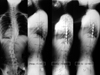
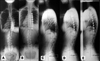
Standing lateral radiographs were analyzed with the Cobb method in the sagittal plane at preoperative, postoperative (within 2 months postoperative), and final follow up (minimum 2 years). The standing lateral radiographs were taken in the position with both arms raised to 90°. The below mentioned radiographic parameters, equivalent to the methods used in the report by Rhee et al.2 were also used. The Measurements included the following: C7 plumbline (the horizontal distance of a plumbline dropped from the center of the C7 body to the posterior-superior corner of the S1 body), proximal junctional measurement (PJM; Cobb angle between the most proximal instrumented vertebra and the segment two levels cephalad), distal junctional measurement (DJM; Cobb angle between the most distal instrumented vertebra and the segment two levels caudal), thoracic kyphosis (T5-T12), and lumbar lordosis (T12-L5). Among above parameters, positive values were used to indicate kyphosis. Radiographs were also analyzed for evidence of instrumentation failure (e.g., broken implants, implant pullout). All radiographic measurements were performed by the third spine surgeon (JO Park), who did not perform VATS.
The basis of pseudoarthrosis was identified as either failed instrumentation or progression of curvature over the instrumented levels. Statistically, by using the repeated measure ANOVA (SPSS 12.0, Seoul, Korea) method, the parameters of preoperative, postoperative, and the last follow up were compared to determine whether they were different.
RESULTS
Preoperative Cobb's angle in coronal plane was 54.5 ± 13.9 and at last follow up it was 19.7 ± 9.3 which improved than preoperatively 34.8° (63.8 % correction).
Fusion levels
Fusion levels varied according to the curve magnitude and level. The average number of instrumented levels were 5.9 levels (5 to 8 levels). The most common proximal level was T5 (25) and the most common distal level was T11 (26). The most proximal fusion level was T4, and the most distal was L1.
Thoracic kyphosis
All patients were relatively hypo-kyphotic before surgery (+18.2 ± 7.7°). There was a 2.2° kyphotic effect for VATS thoracic instrumentation at the immediate postoperative. Over times, this trend became more definite, with a 4.2° increase in thoracic kyphosis at the final follow-up (+18.2 ± 7.7° preoperative to +22.4 ± 7.2° at the last follow-up). The difference in thoracic kyphosis between preoperative and postoperative was 2.2 degree increment in kyphosis (Table 1). The more prominent difference was between preoperative and last follow up (Table 1; p = 0.002). Although the average thoracic kyphotic angle was within normal range, those of 8 cases were less than 20°, the lower limit of the thoracic kyphosis angle. However, there were no clinical symptoms and signs related to hypo-kyphosis (Fig. 1)
C7 sagittal plumbline
All data are shown in Table 1. '+' means anterior displacement as compared with C7 sagittal plumb line. In regard to the entire average of the C7 plumb line, preoperative was -13.9 ± 33.6 mm and finally it was -9.9 ± 23.8 mm. Displacement was on average 4 mm positive (anterior displacement). In the VATS group, the average value of the C7 plumb line was negative preoperative and remained so at the final follow-up (Fig. 2). A statistically significant difference of the C7 plumb line between preoperative and last follow up was not detected (Table 1; p = 0.522).
Proximal junctional measurement
VATS instrumented fusion for the thoracic spine had some changes on the PJM before and after surgery (+6.2 ± 4.3° preoperative and +10.1 ± 4.6° at postoperative). Over times, this trend became obscure. Only a 2.6° increase of PJM (+8.8 ± 3.7 at last follow up) was noticed between preoperative and at the final follow-up. In PJM, the difference between preoperative and postoperative was relatively bigger than the last follow up. However, the difference between preoperative and the last follow up was not statistically significant (Table 1; p = 0.095). Only one patient who underwent VATS instrumentation and fusion had a change in PJM ≥10° at the final follow-up compared with preoperative. However, no patient developed a clinically problematic increase in PJM or underwent revision for that reason.
Distal junctional measurement (DJM)
VATS led to minimal changes in the DJM at the postoperative and the final follow-up compared with preoperative (-6.8 ± 5.1° preoperative to -7.0 ± 3.7° postoperative and -6.7 ± 4° at the last follow-up). Therefore, the significant changes in DJM were not observed from preoperative to the final follow-up. Similarly, in DJM, a significant difference between pre-op and the last follow up was not detected (Table 1; p = 0.88).
Lumbar lordosis
The compensatory lumbar response to selective thoracic anterior instrumentation was maintained in lumbar lordosis after surgery. At the final follow-up, lumbar lordosis was slightly increased (-42 ± 10.7° preoperative to -43.5 ± 11.1° finally). The statistically significant change in lumbar lordosis between preoperative and the last follow up was not detected (Table 1; p = 0.33).
Complications
Four patients experienced atelectasis due to a long operation time. Four patients experienced implant related faults. Of these patients, two had broken caps of the screws in Moss Miami, one had rod failure, and one had a proximal screw pullout. In all 4 cases with implant related faults, clinical symptoms were not detected. Thus, revision surgery was not required. In the case with the rod failure, although pseudoarthrosis was suspected, clinical symptoms were not detected.
DISCUSSION
At a minimum 2 year follow-up, we found that VATS could be used to meet goals of scoliosis surgery similar to the others, previously described surgical techniques.1,2,9 The overall sagittal radio-graphic results, using the VATS, were similar to those of other surgical techniques' results.1-3,5,8
There was a trend for VATS thoracic instrumentation to lead to the postoperative anterior displacement in the C7 sagittal plumbline with a +4 mm increase from -13.9 ± 29.1 mm to -9.9 ± 23.8 mm at the final follow up. The C7 sagittal plumbline that changed in such manners was slightly smaller than the study reported by Rhee et al.2 that reported a 19 mm anterior displacement obtained by open thoracotomy of instrumentation and fusion. However, it was difficult to compare directly because patients were not randomized according to curve type. In addition, in the study by Newton et al.8 was limited to the Lenke type I, the final C7 plumbline obtained was -9 mm by VATS surgery. Although, they did not address the preoperative and operative change in the C7 plumbline, the final C7 plumbline was very similar to that of our study. Even though the positive change of the C7 plumbline between preoperative and at the last follow-up was numerically present, we think that the clinical importance of this finding is negligible. This is because of the low magnitude of the change and the fact that the absolute value of the C7 plumbline at the final follow up remained still negative in the present study.
It has generally gained acceptance with scoliosis surgeons that anterior open surgery is more kyphogenic and posterior surgery more lordogenic. In this study, at the last follow-up thoracic kyphosis was +22.4 ± 7.2°, which was less kyphotic than the average +31° reported by Rhee et al.,2 +30 ± 9° reported by Newton,8 and +33 to +41° reported by Betz et al.5 that were obtained by open thoracotomy. The thoracic kyphosis, +22.4 ± 7.2°, obtained in our study is similar to the result reported by Newton et al.,9 +24 ± 8°, which was obtained in 38 cases of Lenke type I scoliosis by the VATS. The similarities of the study reported by Newton et al.8 and our study are facts that the study population is Lenke type I and the Cobb angle is less than 70°. Although there was no similarity of patients in groups using other surgical techniquess,2,5,8 these results demonstrated the possibility that VATS anterior instrumentation was less kyphogenic over the thoracic spine. Even though, the average thoracic kyphosis at the final follow-up in the VATS group was within the normal range (+20 to +40°), the thoracic kyphosis angle of about one fourth of the cases in this study was below the normal range (+22.4 ± 7.2°). This may be due to the difficulty of the VATS-applying scoliosis surgery for the manipulation of the thoracic kyphosis angle. Although its clinical symptom and significance were not yet detected, we may have more concern to make the normal thoracic kyphosis.
Proximal junctional kyphosis is important as it may affect the functional outcome after posterior spinal fixation surgery, which may be cosmetically unappealing and may induce pain.2,11,12 To assess the development of such proximal junctional kyphosis, Rhee el al. evaluated the surgical outcome by introducing the concept of proximal junctional measurement (PJM) that were the 2 levels of kyphosis angles above the most proximal fusion area.2 In the cases applying open thoracotomy (n = 23), PJM was increased 1° in average; whereas, in the cases applying posterior thoracic instrumentation (n = 40), it was increased by 6°. Thus, they have reported that anterior surgery was effective for the prevention of proximal junctional kyphosis. In the present study, in the VATS instrumented fusion for thoracic spine, it was increased by 2.6°. Thus the result was similar to the open thoracotomy surgical method compared to the posterior surgical method. We were concerned that as VATS generated small thoracic kyphosis among other techniques. Thoracic kyphosis angles were +22.4 ± 7.2° in VATS technique of our series, +24 ± 8° in VATS technique of Newton's series and +30 or more in open thoracotomy series as shown in Table 2.2,5,8 As the compensation, proximal junctional kyphosis may develop; however, it was not different from other studies using open thoracotomy technique. In our study, PJM was increased over 10° in one case after operation. This case was operated at the beginning period of VATS technique and it was very difficult to make enough thoracic kyphosis due to unskilled surgical technique. Although clinical symptom of this case was not found yet, but the sagittal balance of whole spine maintains well. A longer follow-up observation is necessary. In this case, the final PJM was 16°, which was out of normal proximal thoracic kyphosis range (T3-5, 8.5°) defined by Berhardt and Bridwell.13
Since the fixation of VATS to the lumbar spine was not performed, lumbar lordosis may be the secondary effect of thoracic spine fixation. In the studies reporting other VATS outcomes, lumbar lordosis was not mentioned.5,7-11 However, it has been reported that in the group that underwent the instrumentation of the anterior thoracic spine by open thoracotomy, approximately a 5° loss lumbar lordosis was detected immediately after surgery but recovered 2 year after surgery.14 In our study, from 2 months after surgery, it was not different from prior to surgery. After 1 year, it was rather increased by an average 1.5°. VATS also decreased DJM by 0.1°, but it was not considered to be clinically meaningful.
The C7 sagittal plumbline sustained small anterior displacement with VATS thoracic instrumentation, but the overall balance should still remain negative. The change in C7 plumbline was +4 mm and smaller than Rhee's result (post: +9 mm, ant. open: +19 mm)2 (Table 2). The reason of the smaller change in our series seems smaller thoracic kyphosis. Although VATS anterior instrumentation tends to generate less thoracic kyphosis than open thoracotomy technique, there was a little increase of PJM compared to the open thoracotomy surgery, but this parameter belonged to an average normal range. Hence, although the surgical technique of VATS thoracic instrumentation is difficult to make the normal thoracic kyphosis, a acceptable sagittal balance can be obtained in Lenke type I adolescent idiopathic scoliosis. This conclusion is thought to be acceptable because the results of VATS technique locate in the middle of anterior and posterior open surgeries which have been doing frequently with consent.


 XML Download