

 PDF
PDF ePub
ePub Citation
 Print
Print


INTRODUCTION
Following a peripheral nerve injury, low threshold stimuli activate A fibers, which is referred to as the pain state called allodynia. The state of allodynia is one feature of neuropathic pain with complex characteristics and a mechanism that is not completely understood. Histological changes, such as sodium channel activation in peripheral nerves and dorsal root ganglia, reorganization of the primary afferent nerve terminal in the spinal cord, and sympathetic innervation into the dorsal root ganglion, develop following peripheral nerve injury. These are accompanied by functional changes such as peripheral sensitization, central sensitization, and sympathetic excitation resulting in increased hyperexcitability of the central nervous system (CNS) and activation of nociceptive neurons by non-noxious stimuli.1
Clinically, neuropathic pain is resistant to conventional treatment and has both sympathetically maintained pain (SMP) and sympathetic independent pain (SIP) components. Many neuropathic pain animal models have been developed. Injury to tibial and sural nerves is considered a SIP-dominant model, while SMP is prominent in selective spinal nerve ligation model. Systemic administration of phentolamine or guanethidine and surgical lumbar sympathectomy relieved allodynia in the SMP model, but not in the SIP model.2-4
High-voltage-activated calcium channels are heterooligomeric proteins consisting of a poreforming α1 subunit and auxiliary α2δ, β, and in some tissues gamma subunits. Auxiliary subunits support the membrane trafficking of the α1 subunit and modulate the kinetic properties of the channel. In particular, the α2δ subunit has been shown to modify the biophysical and pharmacological properties of the α1 subunit. Alpha2delta subunits can also support pharmacological interactions with drugs that are used for the treatment of epilepsy and neuropathic pain.5,6
Pregabalin binds specifically to the α2δ site, an auxiliary subunit of voltage-dependent calcium channels in the central nervous system. Pregabalin reduces the depolarization-induced calcium influx at nerve terminals, resulting in a reduction of the presynaptic release of excitatory neurotransmitters, including glutamate, noradrenalin, substance P and CGRP.7,8 Pregabalin has a broad analgesic effect on mechanical and thermal hypersensitivity associated with nerve injury, incision injury and inflammatory injury.9,10 Whereas the SMP model has been studied extensively,11-13 the SIP model has not previously evaluated the efficacy of pregabalin against allodynia.
The present study examined and compared the pharmacologic antiallodynic effects of pregabalin on tactile and cold allodynia in SMP- and SIP-dominant rat models of neuropathic pain.
MATERIALS AND METHODS
Experiments were carried out on male Sprague-Dawley rats, with masses ranging from 120-150 g for the SMP model14,15 and 180-220 g for the SIP model.3 Rats were housed in group cages on a 12-h night/day cycle with access to food and water at all times in rooms that were temperature and humidity controlled. The animal surgery and testing procedures were reviewed and approved by the Institutional Animal Care Committee at Yonsei University.
For the SMP model, rats were anesthetized using 3% enflurane in oxygen, and the L6 transverse process on the left side was then partially resected. The L5 and L6 spinal nerves were isolated and ligated tightly using 6.0 black silk just distal to the dorsal root ganglion.14 For the SIP model, rats were anesthetized using 3% enflurane, after which a segment of the sciatic nerve was exposed between the mid-thigh level and the popliteal fossa. The three major divisions of the sciatic nerve (tibial, sural, and common peroneal nerves) were separated. The tibial and sural nerves were ligated using 4.0 black silk and transected, while the common peroneal nerve was left intact.3 For intrathecal (IT) drug administration, a polyethylene (PE-10) catheter was implanted through an incision in the atlantooccipital membrane to the lumbar enlargement after a 7-day postoperative period. The catheter was externalized on the top of the skull and secured with a piece of steel wire. Rats showing postoperative neurological motor deficits were sacrificed immediately.
Behavioral tests for mechanical or cold allodynia on the ipsilateral hindpaw were conducted on all rats on postoperative day 14. Rats were placed in individual transparent acrylic cages with wire mesh floors and allowed to adapt for 30 min.
For the tactile allodynia test, a mechanical threshold was measured by applying 8 calibrated von Frey hairs (0.41-15.1 g) to the sensitive area for 5 sec with enough pressure to slightly bend the hair. Brisk withdrawal or paw flinching was considered a positive response. In the absence of such a response, the filament of next greater force was applied. In the presence of a response, the filament of next lower force was applied. The cutoff value was defined in stimulus intensities of 15 g. The 50% withdrawal threshold was determined using the up-down method.16 Rats with thresholds to the von Frey monofilament less than 3.0 g were included in the study (83% of SMP model rats and 88% of SIP model rats).
Cold allodynia was assessed using the acetone drop application technique.11 An acetone drop was formed at the end of a piece of small polyethylene tubing connected to a syringe. Acetone was applied to the sensitive area of the ipsilateral hindpaw, and each trial of acetone application was repeated five times at intervals of 3 min. Allodynia was assessed by measuring the frequency of foot withdrawal in response to acetone application. Animals that had developed cold allodynia, defined as withdrawal in response to least 4 of the 5 acetone applications, were included (88% in the SMP model and 90% in the SIP model).
Pregabalin (donated from Pfizer Inc, Groton, Connecticut, U.S.A.) was dissolved in 0.9% normal saline, and administered in a volume of 2 mL for IP injection and a volume of 10 µL followed by an additional 10 µL normal saline for IT injection. Dose ranges were 3-30 mg/kg IP and 0.01-100 µg IT to the extent that antiallodynic effect was apparent without sedation or motor impairment (n = 6 in each dose group). Rats were tested 30, 60, 90, 120 and 150 min after IP administration and 15, 30, 45, 60, 75 and 90 min after IT administration. The onset and maximal effect times of IT-administered pregabalin were faster than those of IP-administered pregabalin, so the tests after IT pregabalin administration were performed earlier and more frequently. Motor function was assessed by observing sedation, the placing/stepping reflex and the righting reflex.
Data are presented as the mean ± SEM. The antinociceptive effects of pregabalin are presented as a percentage of the maximum possible effect (%MPE) according to the following formulas: %MPE for tactile allodynia test = (post drug threshold-baseline threshold)/(cutoff threshold-baseline threshold) × 100; %MPE for cold allodynia test = (baseline withdrawal frequency-post drug withdrawal frequency)/(baseline withdrawal frequency) × 100. The ED50 values were statistically analyzed using dose-response analysis, and dose-response data was analyzed using one-way ANOVA.
RESULTS
The majority of rats in both SMP and SIP models exhibited characteristic neuropathic pain behaviors 3 days and 7 days after receiving operations, respectively.
No significant differences were detected in the threshold values for tactile and cold allodynia between the two rat models included in this study after normal saline IP and IT administration (data not shown). The calculated mean tactile threshold values for SMP and SIP rats were 1.84 ± 0.62 g and 1.65 ± 0.71 g, respectively.
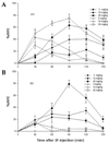
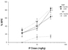
IP administration of 3, 10, 30 mg/kg pregabalin dose-dependently attenuated tactile allodynia in both models. IP-administered pregabalin dose-dependently reduced cold allodynia in the SMP model, but not in the SIP model (Fig. 1 & 2).
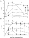
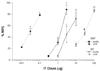
IT-administered pregabalin dose-dependently attenuated both tactile and cold allodynia in both models. However, the dose response curve of IT-administered pregabalin in SMP rats was shifted to the left relative to those of SIP rats, and the ED50 of IT-administered pregabalin for cold allodynia in SMP rats was about 900 times less than that in SIP rats (Fig. 3 & 4).
The maximum suppressive effect on allodynia was reached 30-120 min after IP administration and 15-30 min after IT administration.
No motor impairments were detected in either model following pregabalin administration in the above IP and IT dose range.
DISCUSSION
The present study examined the effect of pregabalin on tactile and cold allodynia in SMP- and SIP-dominant neuropathic pain rat models. We found that selective spinal nerve ligation and tibial and sural nerve transection resulted in similar levels of responses in von Frey filament and acetone drop testing, indicating the development of neuropathic pain in both models. For the tactile allodynia, both IT- and IP-administered pregabalin had antiallodynic effects in both SMP and SIP models. For the cold allodynia, IT-administered pregabalin had much less potent antiallodynic effects in the SIP model compared to the SMP model. And IP-administered pregabalin attenuated cold allodynia in the SMP model but not in the SIP model.
The mechanism of allodynia is complex and is not completely understood. Histological changes in peripheral nerve and dorsal root ganglion, spinal cord, and supraspinal nerve are accompanied by functional changes, such as peripheral and central sensitization and sympathetic excitation, resulting in increased hyperexcitability of the central nervous system and activation of nociceptive neurons by non-noxious stimuli.1 Excitatory amino acids play an important role in the alteration of spinal sensory processing and the plasticity of dorsal horn neurons after damage to the peripheral nervous system.17
While nerve injury-induced mechanical allodynia probably involves sprouting of Aβ-fibers into the superficial dorsal horn and synaptic rearrangement with central sensitization,18 the mechanisms underlying nerve injury-induced cold allodynia have been less well documented. The facilitated responses of C-fibers or membrane property changes in Aδ-cells might be associated with cold allodynia.19-21 Central sensitization mechanisms might also be involved in cold allodynia.18 Thus, the inhibition of cold or mechanical allodynia could result from either the prevention of central sensitization with the direct blockade of noxious stimuli, or from the blockade of low-threshold inputs.22
Autoradiographic analysis of R217A mice shows that the mutation to α2δ-1 substantially reduces specific pregabalin binding in CNS regions that are known to preferentially express the α2δ-1 protein, notably the neocortex, hippocampus, basolateral amygdala and spinal cord. This study provides evidence that the α2δ-1 subunit of voltage-gated calcium channels is the major binding protein for pregabalin in the CNS.23
The upregulation of the spinal dorsal horn calcium channel α2δ subunit contributes to peripheral nerve injury-induced tactile allodynia. Basal expression of the α2δ subunit may occur presynaptically and postsynaptically in the spinal dorsal horn. Nerve injury induces mainly presynaptic α2δ subunit expression that derives from increased amounts of the α2δ subunit in injured DRG neurons. Thus, changes in presynaptic α2δ subunit expression contribute to injury-induced spinal neuroplasticity and central sensitization that underlies neuropathic pain development and maintenance.12,24
Inhibition of excitatory amino acid release from primary afferents by pregabalin binding at calcium channel α2δ subunits could result in reduced availability of glutamate at the NMDA, AMPA and metabotropic receptors.10 A patch clamp study also demonstrated that gabapentin decreased the evoked excitatory postsynaptic current amplitudes mediated by both NMDA and non-NMDA receptors in the superficial lamina, indicating that it had suppressive effects on central sensitization.25 Pregabalin alleviated pain-like behaviors in two (spinal cord injury & sciatic nerve injury) rat models of neuropathic pain, potentially due to the slow releasing kinetics of NO.26 Therefore, ligands bound to α2δ subunits of voltage-dependent calcium channels might be effective at blocking both hyperalgesia and allodynia induced by various thermal and mechanical stimuli.
Luo et al.27 compared DRG and spinal cord α2δ-1 subunit levels and gabapentin sensitivity in allodynic rats with mechanical nerve injuries, a metabolic disorder, or chemical neuropathy. These data indicated that, even though allodynia occurred in all types of nerve injury investigated, DRG and/or spinal cord α2δ subunit up-regulation and gabapentin sensitivity only coexisted in the mechanical and diabetic neuropathies. Thus, induction of the α2δ subunit in the DRG and spinal cord is likely regulated by factors that are specific for individual neuropathies and may contribute to gabapentin-sensitive allodynia. However, the calcium channel α2δ subunit is not the sole molecular change that uniformly characterizes the neuropathic pain states.
While the antinociceptive effect of gabapentin may be produced by spinal, supraspinal, or peripheral inhibitory action on ectopic afferent discharge, the preferential site of antiallodynic action remains the subject of debate.13,28-30 In the present study, IT-administered pregabalin was much more potent than systemic pregabalin in attenuating cold and mechanical allodynia in both models. These observations suggest that the antiallodynic effect of pregabalin is mediated mainly via a spinal mechanism, although supraspinal or peripheral actions cannot be excluded.
The peripheral mechanism of allodynia is mediated by a sodium channel that is blocked with low concentrations of local anesthetic. Expression of auxiliary beta subunits of sodium channel in injured DRG neurons following axotomy is a pathomechanism of post-nerve injury pain in primary sensory neurons.31 However, some studies have shown that pregabalin can reduce the peripheral neurotransmitters, which are the source of the sensitization soup. Pregabalin did not affect the conduction velocity of afferent fibers and the response of normal afferent nerves to mechanical stimulation. These data strongly suggest that the analgesic effect of pregabalin on neuropathic pain is likely mediated, at least in part, by its peripheral inhibitory action on the impulse generation of ectopic discharges caused by nerve injury.30
We could not clearly determine whether the analgesic effect of pregabalin against cold or mechanical allodynia is associated with the sympathetic nervous system. However, sympathetic nerve sprouting into the dorsal root ganglion, especially in SMP, could enhance the pain transmission from the periphery to the spinal cord.32 Thus, we think pregabalin has more potent antiallodynic effects in both tactile and cold allodynia observed in the SMP model. Another possible explanation for the prominent analgesic effect of pregabalin against cold allodynia in SMP is as follows: C-fiber nociceptors acquiring enhanced α-adrenergic sensitivity in SMP might be involved in cold allodynia,20,33 and a spinal role for the voltage-dependent calcium channels in the development of central sensitization is induced by C-fiber activation.34 This suggests that calcium channels might be more responsible for cold allodynia in SMP and that pregabalin could block cold allodynia in SMP more effectively. In SIP rats, IP-administered pregabalin did not block cold allodynia and the analgesic effect of IT-administered pregabalin against cold allodynia was much less potent compared to that in SMP. This suggests less involvement of calcium channels in the mechanism of cold allodynia in SIP.
Large pregabalin doses may reduce the magnitude of motor function. In additional experiments, we found that 100 mg/kg of IP-administered pregabalin caused SIP rats to become sedated and non-ambulatory. In the present study we did not detect any motor impairment, indicating the doses used did not interfere with the animal's ability to respond to acetone or von Frey stimulation.
Both systemic and central pregabalin could treat sympathetically maintained pain. In sympathetic independent pain, central pregabalin has an antiallodynic effect, but systemic pregabalin has no analgesic effect on cold allodynia. The antiallodynic effect of pregabalin is mainly considered to be due to spinal action, and voltage-dependent calcium channels may be less related to the cold allodynia in SIP.
 XML Download