

 PDF
PDF ePub
ePub Citation
 Print
Print


INTRODUCTION
Papillary thyroid carcinoma (PTC) is the most common subtype of thyroid cancer. Although surgery is recommended for PTC, there is no consensus on the therapy when a thyroidectomy is contraindicated because of systemic disease such as heart failure or chronic obstructive pulmonary disease. Hypertrophic cardiomyopathy is a common genetic cardiovascular disease with a high risk of sudden death and disability (1). Given the potential devastating perioperative cardiac complications, hypertrophic cardiomyopathy is generally a contraindication for anesthesia and surgery (2). Radiofrequency ablation (RFA) has been used widely in the treatment of solid cancers (3). There is no report of PTC treated with RFA instead of surgery. Herein, we presented a case of PTC in a patient with hypertrophic cardiomyopathy treated with RFA.
CASE REPORT
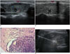
Neck ultrasound performed for a 52-year-old female revealed a predominantly solid nodule (0.9 × 0.7 × 0.5 cm) with marked hypoechogenicty, microcalcification, moderate vascularity, and spiculated margin in the right thyroid gland. Enlarged neck lymph nodes were not detected (Fig. 1A, B). Total thyroxine (TT4) and total triiodothyronine (TT3) were 109.60 nmol/L (55.47–161.25 nmol/L) and 1.16 nmol/L (1.02–2.96 nmol/L); free thyroxine (FT4) and free triiodothyronine (FT3) were 14.63 pmol/L (10.45–24.38 pmol/L) and 4.36 pmol/L (2.77–6.31 pmol/L), respectively. Thyroid-stimulating hormone (TSH) and thyroid peroxidase antibody (TPO-Ab) were 1.58 mIU/L (0.380–4.340 mIU/L) and 852.0 IU/mL (0–100 IU/mL), respectively.
Seven years earlier, the patient had been diagnosed with hypertrophic cardiomyopathy and a DDD cardiac pacemaker was implanted. Her cardiac function was graded as New York Heart Association class III. A cardiac ultrasound showed diffuse thickening of the ventricular walls and the left ventricular ejection fraction was 60%. The N-terminal pro-brain natriuretic peptide was 1048 pg/mL (0–100 pg/mL).
Core needle biopsy (18 G needle) confirmed the diagnosis of PTC (Fig. 1C). The patient was considered unable to tolerate a thyroidectomy, so instead RFA was performed. We used Valleylab Cool-Tip™ RFA System (Boulder, CO, USA), including monopolar electrode, radiofrequency generator and cool-tip pump. The nodule was ablated with fixed electrode technique (FET) (Fig. 1D). After sterilization and local anesthesia with 2% lidocaine, the ablation electrode was inserted into the nodule under ultrasound guidance. The ablation was started with 5 W and increased to 28 W gradually in a 1 cm active tip. It was terminated when the RFA-induced transient hyperechoic zone covered the entire carcinoma and the needle passage was also ablated when the ablation electrode was retracted. It took 7 minutes to complete the ablation. The patient was well informed before RFA was performed.
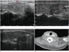
Regular follow-up neck ultrasound showed no signs of recurrence or metastases after 1, 3, 15, and 41 months; and the patient had no complications of RFA (Fig. 2). During the follow-up, the size of ablated lesion was reduced and the echogenicity was decreased; in addition, the TT4 and TT3 were 73.20 nmol/L (55.47–161.25 nmol/L) and 0.94 nmol/L (1.02–2.96 nmol/L), respectively; FT4 and FT3 were 14.02 pmol/L (10.45–24.38 pmol/L) and 3.76 pmol/L (2.77–6.31 pmol/L), respectively. TSH and TPO-Ab were 0.97 mIU/L (0.380–4.340 mIU/L) and 670.70 IU/mL (0–100 IU/mL); respectively.
This case report was approved by the Institutional Review Board.
DISCUSSION
PTC is the most common subtype of thyroid cancer, comprising > 80% malignant thyroid neoplasms. The 10-year survival rate for PTC is 93% (4). Although the prognosis is excellent, the guidelines of the National Comprehensive Cancer Network for thyroid carcinoma and the European Thyroid Association (ETA) both recommend a total thyroidectomy or lobectomy plus isthmusectomy as the standard treatment for PTC (56).
A non-surgical or minimally invasive treatment is required in thyroid cancer patients who cannot undergo surgery due to severe disease, such as hypertrophic cardiomyopathy. As minimally invasive therapy, RFA induces irreversible damage to tumor tissue with heat generated by high-frequency alternating electric current (7). RFA is an established substitute for surgery for solid cancers such as hepatic cancer (3). RFA is also used to treat benign thyroid nodules and metastatic or recurrent well-differentiated thyroid cancer with difficult reoperations (89). The residual volume after RFA is reduced significantly in benign thyroid nodules and metastatic well-differentiated thyroid carcinoma (89). However, whether RFA is a promising therapeutic option for papillary thyroid microcarcinoma (PTMC), which defined as thyroid cancer ≤ 10 mm in diameter, is unclear.
Baek et al. (10) proposed the moving shot technique (MST) for ablation of thyroid nodules by moving the electrode during the procedure. Compared with FET, MST is more suitable for thyroid gland since it can minimize the risk of heat damage to surrounding critical structures such as recurrent laryngeal nerve, blood vessels and esophagus (11). However, at the time, MST was not in use for thyroid nodules in our hospital, and may have been more suitable for this patient.
Papillary thyroid microcarcinoma rarely progress rapidly so the therapeutic intervention remains controversial. Some surgeons recommend active observation as an appropriate strategy instead of immediate surgery. However, the necessity of at least lobectomy for unifocal PTMC is more accepted. ETA and the American Association of Clinical Endocrinologists recommend partial thyroidectomy and lobectomy plus isthmusectomy for PTMC without neck lymph nodes involvement respectively (512). In our case, a calcified 0.9 × 0.7 × 0.5 cm nodule without enlarged lymph nodes was detected and core needle biopsy confirmed papillary thyroid cancer, so surgery was necessary for the patient. Although surgery is the main therapy for PTMC, our case demonstrates that RFA can be a substitute in patients who cannot tolerate surgery. However, neck lymph nodes metastases are quite common for thyroid cancer and central node involvement reportedly exist in 64% of PTMC cases (13). Therefore, for operable thyroid cancer, RFA should be avoided because of undetectable lymph node metastases and patients undergoing RFA as an alternative to surgery should be followed closely. Signs of recurrence or metastases have not been observed on periodical follow-up neck ultrasound in our patient. However, further observation and clinical trials are needed to compare the value of RFA with surgery. RFA may be a reliable therapeutic candidate even for low-risk PTC patients without contraindication of surgery.
In conclusion, to our knowledge, this is the first reported case of PTC with hypertrophic cardiomyopathy treated with RFA. Despite thyroidectomy as prior therapy in our case, RFA is an alternative when surgery is not feasible. However, further long-term observation is needed to confirm the value of RFA in the treatment of thyroid cancer.
 XML Download