

 PDF
PDF ePub
ePub Citation
 Print
Print


INTRODUCTION
Asthma is a common, reversible airway obstructive disease that is characterized by increased bronchial hyperresponsiveness, airway wall inflammation and airway remodeling. CT has been used to assess airway remodeling in patients with asthma. Xenon-inhaled dual-energy (DE) CT has recently been found to be feasible to visualize and quantitate the regional ventilation of the lung (1-5). In asthmatics, the ventilation defects seen on xenon-inhaled DE CT showed significant correlations with the airflow obstruction noted on pulmonary function tests and the airway wall thickening seen on CT (6). We present here the redistributed regional ventilation after the administration of a bronchodilator and this was demonstrated on xenon-inhaled DE CT in a patient with asthma. This case highlights the diagnostic potential of xenon-inhaled DE CT for the evaluation of the regional ventilation changes after the administration of a bronchodilator in patients with asthma.
CASE REPORT
A 7-year-old girl with asthma, allergic rhinitis and atopic dermatitis presented for further evaluation and she underwent a methacholine provocation test as well as pulmonary function tests before and after the administration of a bronchodilator (albuterol sulfate inhalation aerosol). The methacholine provocation test showed that the provocative concentration resulting in a 20% fall in the forced expiratory volume in 1 sec (FEV1), i.e., the PC20, was abnormally low (7.1 mg/mL) and this indicated bronchial hyperresponsiveness (normal bronchial responsiveness: > 16.0 mg/mL). A 17% increase (from 70% to 82%) in the predicted FEV1 and a 112% increase (from 57% to 121%) in the predicted forced mid-expiratory flow rate (FEF25-75%) were recorded after the administration of the bronchodilator and these were regarded as a positive response.
The patient was then recruited for our Institutional Review Board approved study on DE xenon ventilation CT. Written informed consent was obtained from the parents after explaining the potential risks and benefits of DE xenon ventilation CT in this patient. The child was fitted with a face mask (King Systems, Noblesville, IN) and elastic straps to ensure gas-tight delivery of 30% stable xenon (a mixture of 30% xenon and 70% oxygen) with a xenon gas rebreathing system (AZ-726; Anzai Medical, Tokyo, Japan). The DE CT examinations using a dual-source system (SOMATOM Definition; Siemens Healthcare, Forchheim, Germany) were successfully performed twice after inhalation of 30% xenon for 1 min without any side effects, that is, before and 20 min after the administration of the bronchodilator. We decided to add a post-bronchodilator study after we identified multifocal ventilation defects on the xenon-inhaled DE CT and we discussed the potential benefit of the post-bronchodilator study. DE spiral CT scanning of the whole thorax was acquired with 14 × 1.2mm collimation, a 0.45 pitch, a 0.33 second rotation time and a 512 × 512-pixel matrix during end-inspiration breath-holding at the end of 1-min xenon inhalation. The CT dose was reduced to the lowest limit available for the CT system: 15 effective mAs for 140 kV and 64 effective mAs for 80 kV. The volume CT dose index and dose length product were 2.71 mGy and 59 mGy-cm (119 mGy-cm for the two scans), respectively. The total dose estimate of the two DE CT scans was 3.9 mSv. Xenon maps and the weighted-average CT images were generated by using a commercially available workstation (Syngo Dual Energy; Siemens Healthcare, Forchheim, Germany).
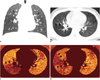
The weighted-average CT images showed diffuse mild wall thickening of the central airways and subsegmental or segmental hyperlucent lung areas that suggested air trapping (Fig. 1). There was no bronchiectasis or mucus plug. The pre-bronchodilator xenon maps revealed a severe segmental ventilation defect in the right lower lobe and a mild subsegmental ventilation defect in the left upper lobe lingular division (Fig. 1C). On the post-bronchodilator xenon maps, the ventilation defect in the right lower lobe was improved, while the xenon values in the right middle lobe slightly dropped and a segmental ventilation defect had newly appeared in the lateral segment (Fig. 1D). On the other hand, a mild subsegmental ventilation defect seen in the left upper lobe lingular division remained unchanged (Fig. 1D).
DISCUSSION
In patients with asthma, ventilation defects are often seen on hyperpolarized gas MRI and the number of defects is correlated with the severity of the disease. The defects may be provoked by the administration of methacholine and they may be improved with the administration of a bronchodilator, which has been demonstrated on hyperpolarized gas MRI. However, hyperpolarized gas MRI is not widely available and there is currently a shortage of helium 3 gas in the world. In contrast, xenon-inhaled DE CT is readily available and it may be used to assess the regional ventilation of the lung (1-6). Chae et al. (6) showed that ventilation defects were seen in asthmatics who had more severe airflow limitation and airway wall thickening, and their extent was correlated with the pulmonary function test results.
The regional ventilation changes after the administration of a bronchodilator and as demonstrated on xenon-inhaled DE CT in this patient may be analogous to those seen on hyperpolarized gas MRI, and they may reflect changes in the regional airflow obstruction that are caused by the inhaled bronchodilator. We think that these changes with a 20 min interval on the same day probably represent bronchodilator responses rather than natural changes over time as the ventilation defects in patients with asthma are unlikely to change during the short time between imaging studies performed on the same day (7). Subsegmental ventilation defects and particularly those in the dependent portions of the lung may be present in healthy subjects. However, the ventilation defects identified in this patient with asthma were obviously larger than those in healthy subjects and they were located in both dependent and non-dependent portions of the lung. In this patient, the most extensive ventilation defect in the right lower lobe was substantially improved after the administration of a bronchodilator, which is an expected finding that indicates a positive bronchodilator response. Of interest, the regional ventilation was conversely diminished in the right middle lobe, which was adjacent to the improved ventilation defect in the right lower lobe. Further study is necessary to clearly define this interesting phenomenon. Nevertheless, it may be speculated that the regional ventilation in the right lung might be redistributed because the improved airflow through the right lower lobe bronchi might unveil that the hidden mild airflow disturbance through the lateral segmental bronchus of the right middle lobe was probably less responsive to a bronchodilator. We also excluded the possibility of a mucus plug for the newly developed ventilation defect seen in the lateral segment of the right middle lobe by confirming the absence of an endobronchial lesion that would suggest a mucus plug in the bronchus, as seen on the multiplanar CT images.
These bronchodilator responses assessed with xenon-inhaled DE CT seemed to reflect those noted on the pulmonary function test in this patient. However, the pulmonary function test provides only the global lung function. Regional ventilation changes after the administration of a bronchodilator can be assessed with hyperpolarized gas MRI. However, the limited availability and a lack of morphologic information about the airway and lung parenchyma are disadvantages of hyperpolarized gas MRI. In contrast, xenon-inhaled DE CT can provide images of both the anatomic and functional lung abnormalities in asthma patients. A previous study (8) also showed that the accurate assessment of airway remodeling and bronchial hyperresponsiveness is an advantage of CT over hyperpolarized gas MRI. Radiation exposure is also a limitation of xenon-inhaled DE CT. However, the CT dose was minimized to the lowest level in this current patient.
In conclusion, changes in the regional ventilation in asthma patients after the administration of a bronchodilator can be demonstrated on xenon-inhaled DE CT.
 XML Download