

 PDF
PDF ePub
ePub Citation
 Print
Print


Tumors arising from the chromaffin cells of the adrenal medulla are called pheochromocytomas, whereas those that occur in the paraganglia at other sites are referred to as paragangliomas (1). Although paragangliomas can be found in every site where healthy paraganglia are known to occur, the majority are commonly seen in the carotid artery, jugular foramen, middle ear, aortopulmonary region, posterior mediastinum and abdominal paraaortic region, including Zuckerkandl's body (2).
Paragangliomas arising from the pancreas are very rare. They have been previously characterized on radiologic images as being well-marginated, hypervascular masses with cystic areas (3-5). However, the reported CT features were those of only single equilibrium phase CT, and this modality can't show the hypervascularity of paragangliomas. Also, the early enhancement of the prominent draining veins during the arterial phase, which supports the hypervascularity of paragangliomas, has not been reported. We herein report on the abdominal ultrasonography (US), dynamic CT and endoscopic retrograde cholangiopancreatography (ERCP) findings that show the extreme hypervascularity of an unusual variant of paraganglioma that arose in the pancreatic head.
CASE REPORT
A 57-year-old woman visited our hospital with right flank discomfort of two-month duration. The patient's past medical history was unremarkable and her blood pressure was normotensive. Her physical examination did not show any abnormality. On admission, the serum CA 19-9 level was slightly elevated at 45.2 U/mL (normal range: 0-37 U/mL). Urine analysis demonstrated three to four white blood cells per high-power field, but the patient denied dysuria or other urinary symptoms. The other laboratory results were within the normal limits.
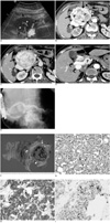
Transabdominal US showed a 6×7-cm, well-marginated, retroperitoneal mass with isoechogenicity relative to the liver parenchyma, and this was adjacent to the pancre atic head. while color Doppler US demonstrated the hypervascularity of the tumor (Fig. 1A). Dynamic pancreas CT revealed a 7-cm, well-demarcated, low attenuating mass in the pancreatic head. On the arterial phase CT, this mass showed strong enhancement and also non-enhancing tubular-shaped portions; several intratumoral vessels that were connected to the peritumoral vessels were also noted. There was early contrast filling of the main portal vein and several prominent draining veins surrounding the mass. On the portal venous phase CT, the mass was still well-enhancing and the extent of the non-enhancing portions within the mass was reduced compared with that seen on the arterial phase. There was diffuse dilatation of the pancreatic duct that was most likely secondary to mass compression or invasion. Given the mass compression of the distal main portal vein, the possibility of main portal vein invasion could not be ruled out (Figs. 1B-D). ERCP demonstrated superior displacement of the head portion of the main pancreatic duct and diffuse mild dilatation of the main pancreatic duct (Fig. 1E). Endoscopic US showed a well-marginated, echogenic mass with several anechoic portions, and this represented cystic degeneration or hemorrhagic necrosis in the pancreas head. It also demonstrated several large vessels within and around the mass and mild dilatation of the main pancreatic duct. Based on these imaging findings, we radiologically diagnosed the tumor as being a non-functioning islet cell tumor of the pancreatic head. We performed pylorus-preserving pancreaticoduodenectomy. The gross specimen revealed a 6.5×6×6-cm, well-circumscribed ovoid soft-tissue mass with multifocal hemorrhagic portions and no cystic degeneration (Fig. 1F). The tumor had displaced the common bile duct and the main pancreatic duct without any evidence of invasion. Microscopic examination showed a Zell-ballen pattern composed of mild pleomorphic chief cells and sustentacular cells within the tumor (Fig. 1G). Prominent peritumoral and intratumoral arteries and veins were also observed. Immunohistochemistrical staining was positive for synaptophysin, chromogranin and S-100 protein, but negative for cytokeratin (Figs. 1H, I). The histopathologic findings were consistent with a paraganglioma of the pancreas.
DISCUSSION
Paragangliomas or extraadrenal pheochromocytomas are rare, affecting about one in 2,000,000 people (6). Although most paragangliomas are solitary and they arise sporadically, they can be multicentric or hereditary. Paragangliomas of the abdomen predominantly arise from paraganglia that are symmetrically distributed along the abdominal aorta in the retroperitoneum. The most prominent collection is near the origin of the inferior mesenteric artery (the organ of Zuckerkandl), which is where the majority of abdominal paragangliomas originate. Other less common locations of abdominal paragangliomas include the gallbladder, urinary bladder, prostate, spermatic cord, uterus and duodenum (2). Although paragangliomas occur in a variety of anatomic locations, they have nearly identical imaging features, namely a homogeneously or heterogeneously hyperenhancing, soft-tissue mass with cystic areas on CT scanning and multiple areas of signal void interspersed with hyperintense foci (a salt-and-pepper appearance) within the tumor on the MR imaging (2).
Paragangliomas of the pancreas are very rare. To the best of our knowledge, only eight cases of pancreas paraganglioma have been reported (3-5). The mean age of these eight patients was 67 years (range: 42 to 85 years) with a male to female ratio of 1/7. Six of these eight tumors were located in the pancreatic head, whereas the remaining two originated from the body of the pancreas. Although the available radiologic images were limited in the previously reported cases of pancreas paraganglioma (3-5), the imaging findings were generally characterized as a well-defined mass with frequent areas of hypoechogenicity on US, a well-marginated, hypervascular tumor with cystic areas of low-attenuation on contrast-enhanced CT, and tumor displacement of the main pancreatic duct on ERCP. Although the present case is characterized by similar findings, it is unique because the dynamic CT demonstrated robust enhancement of the mass that was comparable to that of the greater abdominal vessels, prominent intratumoral vessels and early contrast filling of the main portal vein and draining veins from the mass during the arterial phase.
The differential diagnosis of a hypervascular pancreatic mass should include islet cell tumor (ICT), which can be functioning or nonfunctioning according to their clinical and laboratory manifestations (7). Functioning ICTs are usually less than 3 cm in size and they are homogeneously hyperenhancing during the arterial phase of contrast-enhanced CT (7). On the other hand, nonfunctioning ICTs tend to be larger than functioning ICTs, they have a greater predilection for cystic change or necrosis, and they are heterogeneously enhancing (7). The radiologic differentiation of pancreas paragangliomas from nonfunctioning ICTs can be difficult. Yet to the best of our knowledge, there is no report regarding early contrast filling of the prominent draining veins of nonfunctioning ICTs. Therefore, early contrast filling of the prominent draining veins of this tumor and the portal vein too may be a clue for differentiating pancreas paragangliomas from nonfunctioning ICTs of the pancreas.
In conclusion, we report here on a patient with a rare diagnosis of primary paraganglioma of the pancreas, and this tumor was characterized by hypervascularity with prominent intratumoral vessels and early contrast filling of the draining veins from the mass. Despite its rarity, paragangliomas should be a part of the differential diagnosis of a hypervascular pancreatic mass, and especially in the setting of early contrast filling of the prominent draining veins from the mass.
 XML Download