

 PDF
PDF ePub
ePub Citation
 Print
Print


Clonorchiasis is a snail-transmitted, parasitic disease of the bile ducts; this is caused by chronic infestation of liver flukes, Clonorchis sinensis, which reside mainly in the medium- and small-sized intrahepatic bile ducts (1-4). The CT, ultrasonograms and cholangiograms of clonorchiasis patients usually show diffuse, uniform, minimal or mild dilatation of the small intrahepatic bile ducts, particularly in the periphery, without dilatation of the extrahepatic bile duct (1-4). Clonorchiasis is one of the most frequent causes of cholangiocarcinoma and cholangitis in the endemic countries; therefore, its early diagnosis is critical in order to prevent complications such as cholangiocarcinoma, biliary stones and cholangitis (4). We report here on the CT findings of an unusual case of hepatic parasitic abscess caused by clonorchiasis; this malady mimicked cholangiocarcinoma, and there was no dilatation of the intrahepatic bile ducts.
CASE REPORT
A 52-year-old man was referred to our hospital with a liver mass that had been incidentally detected on a liver CT at an outside hospital. He presented with general weakness of one month duration. He denied a past history of eating raw freshwater fish. The results of his physical examination were unremarkable. The results of his laboratory tests, including the peripheral eosinophil count, serum alkaline phosphatase, serum bilirubin, aspartate aminotransferase and alanine aminotransferase, were all in the normal ranges. The viral markers for hepatitis were all negative. The levels of tumor markers, including α-fetoprotein, CA 19-9 and carcinoembryonic antigen (CEA), were normal. His past medical history was unremarkable except for laparoscopic cholecystectomy that was performed due to a gall stone six years previously.
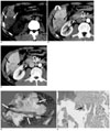
In our hospital, this patient underwent two-phase liver dynamic CT because the CT images taken outside our hospital were not available. The CT showed a 4-cm, illmargined, lobulated, low-attenuation mass in the right posteroinferior segment of the liver (Figs. 1A-C). The mass had heterogeneous, peripheral contrast enhancement during the hepatic arterial phase and then more central enhancement was seen from the peripheral portion of the mass during the portal venous phase (Figs. 1B, C). A transient hepatic attenuation difference that surrounded the mass was noted during the hepatic arterial phase. There was a small portion of poorly enhancing, low attenuation in the peripheral area of the mass, which represented a necrotic portion. Neither the intrahepatic nor extrahepatic bile ducts were dilated. There was an approximately 1.5-cm sized lymph node in the aortocaval space (Fig. 1C). The differential diagnosis of the mass was cholangiocarcinoma or abscess. The patient did not undergo percutaneous biopsy because he had little clinical evidence of a hepatic abscess. The patient underwent segmentectomy of the liver and resection of the lymphadenopathy in the aortocaval space. Pathologic examination showed an ill-defined, lobulated mass measuring 7×6×5 cm (Fig. 1D). On histologic examination (Fig. 1E), the mass consisted of inflammatory cells such as lymphocytes, neutrophils and macrophages, and also granulation tissue. A granulomatous reaction was also seen in the mass. A large number of parasitic eggs were observed among the inflammatory cells. The histopathologic diagnosis was parasitic abscess caused by clonorchiasis along with a granulomatous reaction. The abscess involved the liver resection margin and the hepatic capsule. The lymphadenopathy was considered to be reactive hyperplasia. The patient took antiparasitic medication for the next two months. One year after his discharge, the follow-up CT showed no unusual findings except for segmentectomy of the liver.
DISCUSSION
Clonorchiasis is widely distributed in Asia, from Japan to Vietnam. Although the incidence of human clonorchiasis infections has gradually decreased, it is still estimated that about 15 million people worldwide are currently infected (1). Human infestation depends on the eating habit of repeatedly ingesting raw freshwater fish. The clinical manifestations depend on the number of flukes, the period of infestation and the complications such as pericholangitic abscess, recurrent pyogenic cholangitis, bile duct stones and cholangiocarcinoma (1).
The radiologic findings of clonorchiasis are well known. On CT, the typical findings of clonorchiasis are uniform, minimal or mild dilatation of the intrahepatic bile ducts without dilatation of the extrahepatic bile ducts or any focal obstructive lesions (1-4). The peripheral intrahepatic bile ducts are obstructed by numerous parasites that measure 8-15 mm in length and 1.5-4 mm in width, and so the intrahepatic bile ducts become dilated. However, the extrahepatic bile duct doesn't become dilated because it is not easily obstructed by the parasites. There are many other conditions that can occlude the intrahepatic bile ducts, including adenomatous hyperplasia, mucus, periductal fibrosis and stricture (4). Several investigators (2, 3, 5) have reported that there was no appreciable dilatation of the intrahepatic bile ducts on the CT scans of clonorchiasis patients, but they didn't present any reasons for the absence of dilation. The intrahepatic bile ducts in our patient were not dilated on CT scans, making the preoperative diagnosis of clonorchiasis very difficult. Although our patient did not recall any history of raw or partially cooked freshwater fish, we presumed that acute infestation of clonorchiasis caused no appreciable dilatation of the intrahepatic bile ducts on CT. Pyogenic hepatic abscess is one of the complications of clonorchiasis. Choi et al. (3) have reported a case of multiple small pyogenic abscesses with diffuse dilatation of the intrahepatic bile ducts. Mirdha et al. (6) have also described acute clonorchiasis in a child who had presented with multiple hepatic abscesses and mild dilatation of the biliary tree. To the best of our knowledge, this is the first case in the English medical literature of a hepatic parasitic abscess caused by clonorchiasis without any perceptible dilatation of the intrahepatic bile ducts, as seen on CT scans. Seong et al. (7) reported that the following CT findings favor hepatic abscess over the mass-forming type intrahepatic cholangiocarcinoma: multilayered enhancement, a sharp margin, inner air-density, a cluster sign, an air-biliary gram, a lobulated configuration, atelectasis of the lower lungs, pleural effusion and transient hepatic attenuation difference. In daily practice, however, such differentiation poses difficulty given the considerable overlap of the CT findings. In the present case, the CT findings suggestive of cholangiocarcinoma (i.e. peripheral enhancement of the mass and lymphadenopathy) and the absence of any clinical or radiological findings of a hepatic abscess caused great difficulty in making a preoperative diagnosis of hepatic abscess.
In summary, we report here on a rare case of a hepatic parasitic abscess that was caused by clonorchiasis without dilatation of the intrahepatic ducts, and this malady mimicked cholangiocarcinoma on the CT images.
 XML Download