

 PDF
PDF ePub
ePub Citation
 Print
Print


Osteomas are benign neoplasms consisting of mature normal osseous tissue. A common location is the long bones of the extremities; in the head and neck region they are found in the sinuses, facial bones, skull and mandible (1-5). A subdural osteoma is extremely rare: to the best of our knowledge, it has been described in only two English-language case reports (2, 3). In this article, we report the CT and clinicopathologic findings of subdural osteoma.
CASE REPORT
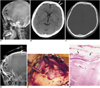
A 43-year-old woman presented with a two-year history of constant headache, unrelieved by analgesics, in the left frontal area. She had no history of head trauma, and physical examination revealed no neurological or systemic abnormality. Routine laboratory tests were within normal limits. Skull radiography, however, revealed the presence of an ovoid radiopaque lesion in the left frontal area (Fig. 1A), and computed tomography (CT) depicted a lentiform ossified lesion in the left frontal skull, obliterating the adjacent CSF space (Fig. 1B). At a bone window width/level setting of 2500/200, a curvilinear lucent line was noted between the inner table of the skull and the ossified mass (Fig. 1C). The preoperative diagnosis was intraosseous osteoma, and for surface marking, plain radiography was used.
Left frontal craniotomy failed to reveal a nodular elevated lesion at the inner table of the skull. The surface of the dura mater at the craniotomy site was smoothly elevated, however, and intraoperative radiography showed that the cause of this was an ossified lesion (Fig. 1D). The dura mater was opened and reflected, and a whitish, stony, nodular lesion was found in the subdural space. It was firmly attached to the inner surface of the dura (Fig. 1E), compressing the underlying brain, but not adhering to the arachnoid membrane. It was removed as one piece, together with adherent overlying dura, and was found to measure 1.2×2.0×0.7 cm. The dural defect was closed using an artificial dural substitute. Histologically, the lesion consisted of mature lamella bone, made of Haver's system, and normal osteocytes between osteoid layers (Fig. 1F).
The patient recovered without complication, and at follow-up six months later, was asymptomatic.
DISCUSSION
The predilective sites of intracranial osteomas are the periosteum of the frontal or ethmoid sinuses, and the inner table of the skull, for which the dura functions as the periosteum: it has been postulated that these neoplasms arise as new bone is formed from the dura or falx (1, 2). An intracranial osteoma can be confused with the dural metaplastic ossification commonly seen in elderly individuals: Fallon et al. reported that at the time of death, up to 5% of adults have meningeal osteomas, and that the radiological findings at this time usually indicate the presence of falx calcification (1). A basis for differentiation between dural osteoma and metaplastic dural ossifications is that the latter are commonly multicentric and form plaque-like or nodular deposits. They are generally located on both sides of the dural falx junction along the superior sagittal sinus and manifest as incidental radiological or postmortem findings (1, 2). A subdural osteoma is a rare lesion. It has a wide base and arises from the inner layer of the dura mater, growing inward as an expanding mass with a well-outlined border and causing pressure symptoms by compressing or displacing the underlying brain. The persistent headache experienced by our patient may have been due to irritation or compression of the adjacent dura, and was cured by surgical removal of the subdural osteoma (2, 3).
Radiological studies may be useful in demonstrating the radiopaque nature of an osteoma. CT examination is sometimes sufficient for the diagnosis of osteoma of the inner table of the skull, in which a dense, mushroom-like lesion is attached to that area by a bony stalk or neck (6). Routine CT is, however, an unreliable basis for definitive differentiation between a subdural and an 'ordinary' intraosseous osteoma. In our case, the lucent line seen at the bone window setting was a clue that suggested a subdural or intradural location. The line, which appeared to represent intervening dura between the skull and subdural osteoma, is considered to be one of the differential points between osteoma of the inner table of the skull and meningioma (6). With the application of magnetic resonance (MR) imaging, the nature and site of attachment of subdural or intradural lesions may readily be discerned (2).
In conclusion, detailed CT examination can provide precise information about the origin of an ossifying lesion in extra-axial space. If CT scanning reveals a lucent dural line, this may indicate that the lesion has an intradural or subdural origin.
 XML Download