

 PDF
PDF ePub
ePub Citation
 Print
Print


INTRODUCTION
Castleman disease is a rare benign lymphoproliferative disorder that causes progressive enlargement of lymph nodes (LN). The histopathogenetic classification of Castleman disease distinguishes hyaline vascular, plasma cell, and human herpesvirus 8-associated Castleman disease, and multicentric Castleman disease not otherwise specified (1). While Castleman disease most commonly occurs in the mediastinum, head and neck involvement accounts for 15-20% of all cases. Although it most commonly involves cervical level I, II, and III LN in the head and neck, involvement of level IV, V, supraclavicular and intraparotid LN also has been reported (2-4). However, to the best of our knowledge, imaging features of Castleman disease involving infrathyroidal region have not been presented in the literature. Infrathyroidal Castleman disease may pose a diagnostic dilemma because it may mimic more common parathyroid adenoma as well as nodal diseases, such as lymphoma. We report here a case of Castleman disease of hyaline vascular type of the infrathyroidal paratracheal region (level VI) in a 48-year-old woman, along with its CT and sonographic findings. Histological features of the specimen were obtained by sonographically guided core-needle biopsy (US-CNB) and surgical excision.
CASE REPORT
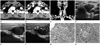
A 48-year-old woman was referred to the department of radiology for further evaluation of the left infrathyroidal mass incidentally, which was identified during sonographic screening of the thyroid gland at a local clinic. Her physical examination and past history were unremarkable. There was no palpable mass in the left infrathyroidal region or elsewhere in the head and neck regions. Serum parathyroid hormone level was 61 pg/mL (reference range, 13-54 pg/mL). Other laboratory findings, including serum calcium and phosphorus levels, were within the normal limit. Multidetector CT performed by using a Sensation 64 scanner (Siemens, Erlangen, Germany) showed an approximately 22 × 21 × 31-mm-sized mass in the left infrathyroidal paratracheal region (level VI) (Fig. 1A-C). The mass was well-demarcated, ovoid, and slightly lobular (Fig. 1A-C). It was nearly isoattenuating to the adjacent muscles on non-enhanced CT (NECT) images (Fig. 1A), and was homogeneous and intensely enhanced on contrast-enhanced CT (CECT) images (Fig. 1B, C). Sonographic examination, which was performed using an Acuson Sequoia 512 scanner (Siemens Medical Solutions, Mountain View, CA, USA) equipped with a 8- to 15-MHz linear array transducer, revealed a well-demarcated, slightly lobular, homogeneous, and markedly hypoechoic (more hypoechoic compared with the adjacent muscles) mass with prominent central and peripheral vascularity on gray-scale and power Doppler (PD) ultrasonography (US) (Fig. 1D-F). In view of CT and sonographic features, our tentative diagnosis was parathyroid adenoma and lymphoma. The surgeon requested US-CNB for histological diagnosis. Due to intense enhancement and prominent vascularity on CECT and PD US, we cautiously performed US-CNB by using a freehand technique with a 18-gauge needle (Bard Peripheral Technologies, Covington, GA, USA) and a spring-loaded, single-action biopsy gun (Pro-Mag 2.2, Manan Medical Products, Northbrook, IL, USA) with an excursion length of 15 mm. Three samples were obtained from the mass for histological examination and immunohistochemical study. No evidence of hemorrhage was noted within and around the mass on sonographic monitoring during and immediately after the procedure. With application of manual compression of the lesion for approximately 20 minutes, the patient experienced no significant bleeding after the procedure. The histological examination of the specimen obtained by US-CNB demonstrated numerous small lymphocytes and markedly increased vascularity without germinal centers (Fig. 1G), whereas immunohistochemical staining for CD20 revealed strong positivity for numerous small B cells (Fig. 1G). The histological and immunohistological findings of US-CNB specimen were interpreted as lymphoproliferative lesion, and thus were inconclusive. She underwent surgical excision of the mass. The histological examination of the specimen obtained by surgical excision exhibited germinal centers pierced by a hyalinized blood vessel ("lollipop" appearance) and were cuffed by the characteristically expanded mantle zone composed of small lymphocytes arranged in a concentric onionskin pattern, which were characteristics of Castleman disease of hyaline vascular type (Fig. 1H).
DISCUSSION
Hyaline vascular type accounts for 90% of Castleman disease's cases and is unicentric in 90% of the cases. It usually manifests as an asymptomatic mass lesion. On the other hand, plasma cell type accounts for less than 10% of Castleman disease's cases, and is multicentric in 76-91% of the cases. It is frequently associated with systemic manifestations, such as fever, night sweats, malaise, hematologic and immunologic abnormalities, such as anemia, thrombocytopenia, and hyperglobulinemia, and splenomegaly (1, 5). Microscopically, the hyaline vascular type is characterized by abnormal small follicles and interfollicular vascularity consisting of a network of small capillaries with a thickened, hyalinized wall penetrating the germinal centers ("lollipop" appearance), which are cuffed by the characteristically expanded mantle zone composed of small lymphocytes arranged in a concentric onionskin pattern.
The clinical diagnosis of cervical Castleman disease of hyaline vascular type is difficult because most patients are present only with a painless cervical mass and there are no laboratory abnormalities suggesting this entity. The classic CT feature of hyaline vascular Castleman disease is that of a solitary enlarged lymph node, presenting homogeneous, intense enhancement after contrast material administration. Unfortunately, the classic CT feature is nonspecific and can be seen in other entities, such as lymphoma, metastatic papillary thyroid carcinoma, schwannoma, and myopericytoma. As in our case, infrathyroidal Castleman disease might pose further diagnostic dilemma, given the high likelihood of parathyroid adenoma in this location, in addition to the intense enhancement of parathyroid adenoma on CECT. In contrast to the classic CT feature of homogeneous, intense enhancement, lesions of Castleman disease of hyaline vascular type with mild to moderate and heterogeneous enhancements have also been reported (2, 4, 6). Glazer et al. (7) have proposed that central stellate hypointensities seen within the cervical LN on T2-weighted MR images, which might have been attributed to calcification, fibrous septations, or vessels in the enlarged LN of hyaline vascular Castleman disease, may help narrow the diagnostic possibilities (7). Tan et al. (6) noticed a central non-enhancing area in the enhancing LN in two of three cases with Castleman disease of hyaline vascular type on CECT. It was crescentic or stellate and corresponded to the fibrous scar on gross pathologic examination. They proposed that the presence of a central non-enhancing scar in an enhancing LN in the neck on CECT could be an important diagnostic clue of hyaline vascular Castleman disease.
Sonographic features of Castleman disease of the head and neck have rarely been presented in the literature (8). Sonographic features of all presented cases of hyaline vascular type were well-demarcated, homogeneous, hypoechoic mass with increased vascularity in both central and peripheral portions (8, 9). They are nonspecific and can be seen in other entities, including lymphoma. Meanwhile, parathyroid adenoma usually appears as a round or ovoid, homogeneous, hypoechoic mass with increased vascularity (10). As such, infrathyroidal Castleman disease of hyaline vascular type may also masquerade parathyroid adenoma on US, as in our case. Similarly, on rare occasions, lymphoma may occur as an infrathyroidal solitary nodal mass with homogeneous enhancement on CECT, along with marked hypoechogenicity and increased vascularity on US, which may mimic parathyroid adenoma and Castleman disease of hyaline vascular type. In view of CT and sonographic findings, we suggested parathyroid adenoma and lymphoma as a possible diagnosis. We could not assume Castleman disease preoperatively due to its relative rarity.
US-CNB of the head and neck lesion is a safe, effective, and time-efficient procedure if performed by an experienced physician. The use of coaxial guiding needle might diminish the tissue injury. Furthermore, immunohistochemical examination of the specimens obtained by US-CNB will enhance diagnostic accuracy. However, the results of histological and immunohistochemical examination of the specimen obtained by US-CNB in our case suggested lymphoproliferative lesion; thus, it was considered to be of limited value for the definitive diagnosis of Castleman disease of hyaline vascular type.
In summary, we suggest Castleman disease of hyaline vascular type as an alternative diagnostic possibility for a well-demarcated infrathyroidal paratracheal (level VI) mass with homogeneous, intense enhancement on CT, and with marked hypoechogenicity and hypervascularity on gray-scale and PD US. Hence, parathyroid adenoma and lymphoma should be included in the differential diagnosis. US-CNB seems to be of limited value in the histological diagnosis of Castleman disease of hyaline vascular type.
 XML Download