

 PDF
PDF ePub
ePub Citation
 Print
Print


Abstract
Cerebral air embolisms generally result from invasive procedures such as a percutaneous needle biopsy, chest tube insertion, central venous catheter access or removal, operations and so on. Likewise, they are mostly iatrogenically induced and present various degrees of severity depending on the number of air bubbles. With the exception of divers, the occurrence of a cerebral air embolism in the absence of invasive procedures is very rare. We report a case of a cerebral air embolism that developed following defecation and was detected by CT in a patient with extensive pulmonary tuberculosis.
Figures and Tables
Fig. 1
A. Chest X-ray shows advanced pulmonary tuberculosis.
B. A left internal mammary angiography on the day of admission reveals extensive hypervascularity in the left lung with an arteriovenous fistula from the left internal mammary artery (white arrow) to the pulmonary vein (black arrow).
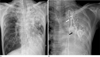
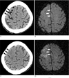
Fig. 2
A. Non-contrast axial CT demonstrates curvilinear low attenuation lesions in the sulci of the right frontal lobe (black arrows), suggestive of air embolism, 10 minutes after onset of the stroke.
B. Diffusion-weighted MR image obtained 3 hour after onset of the stroke shows no abnormal high signal intensity at right frontal lobe.
C. 2 days after the stroke, the air bubbles have disappeared and the cerebral sulcus and cortex (white arrows) becomes effaced and swollen at the same area on the CT scan.
D. Diffusion-weighted MR image shows progression of the cortical high-intensity indicating acute infarction in the same area.
References
1. Kau T, Rabitsch E, Celedin S, Habernig SM, Weber JR, Hausegger KA. When coughing can cause stroke-a case-based update on cerebral air embolism complicating biopsy of the lung. Cardiovasc Intervent Radiol. 2008; 31:848–853.
2. Yamashita Y, Mukaida H, Hirabayashi N, Takiyama W. Cerebral air embolism after intrathoracic anti-cancer drug administration. Ann Thorac Surg. 2006; 82:1121–1123.
3. Hsi DH, Thompson TN, Fruchter A, Collins MS, Lieberg OU, Boepple H. Simultaneous coronary and cerebral air embolism after CT-guided core needle biopsy of the lung. Tex Heart Inst J. 2008; 35:472–474.
4. Muth CM, Shank ES. Gas embolism. N Engl J Med. 2000; 342:476–482.
5. Helps SC, Parsons DW, Reilly PL, Gorman DF. The effect of gas emboli on rabbit cerebral blood flow. Stroke. 1990; 21:94–99.
6. Murphy BP, Harford FJ, Cramer FS. Cerebral air embolism resulting from invasive medical procedures. Treatment with hyperbaric oxygen. Ann Surg. 1985; 201:242–245.
7. Ashizawa K. Possible airflow around the needle in lung biopsy. AJR Am J Roentgenol. 2005; 185:553.
 XML Download