

 PDF
PDF ePub
ePub Citation
 Print
Print


INTRODUCTION
The use of laparoscopic or robotic surgeries for recto-sigmoid colon cancer, prostate cancer, and diseases of the female genital organs in the pelvic cavity is increasing. These surgeries are typically performed in a steep Trendelenburg position, which increases the intraocular pressure (IOP) by 13‑26 mmHg compared with the preoperative IOP value (12). The incidence of ophthalmological complications has not yet been clearly reported after surgeries in Trendelenburg position (12). Although rare, postoperative ophthalmological complications should be considered in patients receiving surgeries in Trendelenburg position since it is destructive and distressing once it occurs. Weber et al. (3) reported that posterior ischemic optic neuropathy developed after a minimally invasive prostatectomy using a da Vinci robot system in a steep Trendelenburg position. This has been attributed to the fact that the ophthalmic circulatory autoregulation does not work properly under general anesthesia, and the ocular perfusion pressure (OPP; mean arterial pressure (MAP) minus IOP) decreases continuously as a consequence (2).
Apart from the surgical position, hypertension is another factor that influences the IOP. The relationship between hypertension and IOP has been established in animal studies, and hypertension is one of the major causes of glaucoma (4). However, no concrete relationship has been validated between hypertension and IOP under particular circumstances such as surgery and general anesthesia. It is meaningful to compare the changes in IOP between normotensive patients and hypertensive patients during surgery in a steep Trendelenburg position because the prevalence of hypertension is increasing and the frequency of surgery in hypertensive patients is thus growing.
There have been few studies on attenuation of the increase in IOP and maintenance of OPP during surgery in a steep Trendelenburg position. Topical application of brimonidine, an α-2 agonist, before general anesthesia resulted in a slight decrease in the intraoperative time-weighted average IOP, by 4 mmHg, and a bolus injection of gabapentin or dexmedetomidine as a premedication before tracheal intubation alleviated the increase in IOP (567). The most important factor that causes increased IOP in a steep Trendelenburg position is increased episcleral venous pressure (8). An α-2 agonist reduces IOP by increasing the uveoscleral outflow and reducing aqueous production (9). Dexmedetomidine, a potent α-2 agonist, would thus seem to be an effective agent for preventing the IOP increase and maintaining the OPP when a patient is in a steep Trendelenburg position. Moreover, as this agent has a short terminal half life (~2 hours) relative to the longer total surgery time, it may be more effective to infuse the α-2 agonist continuously than to administer it as a bolus, so that its effect lasts throughout the period of the steep Trendelenburg position.
Thus, we designed this study to assess whether continuous infusion of dexmedetomidine has any beneficial effect on changes in the IOP and OPP in patients undergoing laparoscopic or robotic surgery in a steep Trendelenburg position. We also evaluated the effects of underlying hypertension on the IOP, compared with normotensive patients.
MATERIALS AND METHODS
Study population and ethical approval
Adult patients with an ASA physical status of class I or II, and who were scheduled for elective laparoscopic or robot-assisted surgery due to recto-sigmoid colon cancer, prostate cancer, or gynecological cancer between March and June 2015, were enrolled in this randomized, placebo-controlled, prospective study. Patients with previous eye surgery, allergy to the study drug, preexisting eye disease including glaucoma, or preoperative unstable hemodynamics were excluded.
Study protocol
When patients arrived in the operating theater, they were allocated to the dexmedetomidine or saline group using block randomization. Basic monitoring, including ECG, noninvasive blood pressure, pulse oximetry, and bispectral index (BIS), was applied, and an IOP measuring device was prepared. In the dexmedetomidine group, 1.0 µg/kg of dexmedetomidine was administered before induction of general anesthesia, and 0.5 µg/kg/hr of dexmedetomidine was infused continuously after tracheal intubation. In the saline group, the same volume of saline was administered in an identical way as in the dexmedetomidine group. An anesthesiologist not involved in the anesthetic management of the patients prepared the covered syringe pump for dexmedetomidine and placebo solutions and held the randomization codes until the end of the study. Another anesthesiologist who was not involved in any way with perioperative patient evaluation and did not know which drug was assigned conducted the entire course of anesthesia. Both the patients and the anesthesiologist in charge were blinded to the group allocation for the duration of the study.
For anesthesia induction, 1.5 mg/kg propofol and 0.8 mg/kg rocuronium were administered and the trachea was intubated after 90 seconds of manual ventilation with 100% oxygen and 3-4 vol% sevoflurane. The right internal jugular vein was catheterized for fluid management and measurement of central venous pressure (CVP). Anesthesia was maintained with sevoflurane 1.5-2.0 vol% and remifentanil 60-300 µg/hr, keeping the BIS at ~50. Mechanical ventilation was maintained using 50% air and 50% oxygen, with an end-tidal carbon dioxide (EtCO2) of 30-35 mmHg. A target hemoglobin concentration of 10-12 g/dL, a CVP of 10 mmHg, and a MAP between ± 20% of the baseline value were attained with appropriate fluid management (3-5 mL/kg/hr) and transfusion at the discretion of the anesthesiologist. Normosol-R was used for crystalloid solutions. For blood loss below 5% of the estimated blood volume, an equal volume of 6% hydroxyethyl starch was administered, and packed red cells were transfused for blood loss of over 5% of estimated blood volume.
Pneumoperitoneum was created by intraperitoneal insufflation with CO2 while the patient was in the supine position. Patients were then placed in the steep Trendelenburg position (30°‑35°). All operations were performed at the same angle on the same table (Alphastar 1132.01 A/B, Maquet GmbH & Co., Wayne, NJ, USA). Throughout surgery, the intraperitoneal pressure was maintained at 15 mmHg, using CO2 for insufflation.
After applying two drops of 0.5% proparacaine HCl (S.A. Alcon-Couvreur N.V., Puurs, Belgium) for topical anesthesia, the IOP was measured 16 times: before anesthetic induction (baseline value, T1); before administration of the study drug (T2); after administration of anesthetic induction agents (T3); after tracheal intubation (T4); 1, 3, 5, and 10 minutes after tracheal intubation (T5-T8); immediately after intraperitoneal CO2 insufflation (T9); immediately after the steep Trendelenburg position (T10); 1, 2, and 4 hours after the steep Trendelenburg position (T11-T13); just before the supine position (T14); and 10 and 30 minutes after the supine position (T15, T16). The IOP was measured with a hand-held tonometer, (Tono-Pen AVIA, Reichert Technologies, Depew, NY, USA). The tonometer averages five successful readings and displays the mean with 95% confidence intervals. Measurements were retaken if the range was greater than 5%. At the time of each tonometer reading, the following data set was collected: MAP, heart rate, EtCO2, peak inspiratory pressure (PIP), and mean inspiratory pressure (Pmean). All fluid and blood products administered were recorded, blood loss was estimated, and urine output was measured. The length of time in the steep Trendelenburg position was noted. The total amount of remifentanil administered was also recorded. In the recovery room, patients were asked about any vision change or eye discomfort. The OPP was calculated as MAP minus IOP. After completion of data collection, the patients in each group were subdivided into two groups according to the presence of underlying hypertension, and the IOP at each time point was compared between the normotensive and hypertensive patients.
Statistical analysis
For sample size calculation, a t-test was performed to evaluate differences in the IOP at 1 hour in the steep Trendelenburg position between the dexmedetomidine and saline groups in a pilot study. The Δ [Δ =|u2 - u1|/σ] of the IOP value was 0.8, with a 6 mmHg difference in mean values between groups and a standard deviation of 7.5. The sample size required at a level of significance of 5% (2-sided α = 0.05) and a power of 80% (1-β = 0.8) was 26 patients per group. All data were analyzed using SPSS software (ver. 18.0; SPSS, Inc., Chicago, IL, USA). To compare demographic data, a χ2 test and t-test were used. Repeated-measures ANOVA was performed to compared the IOP, OPP, PIP, and Pmean between the two groups; with ‘group’ and ‘time point’ as independent variable, after confirming normal distribution with the Shapiro-Wilk test (P > 0.05). The interaction term was calculated with Bonferroni correction for repeated measures. The relationships between the PIP or Pmean and the IOP were analyzed using Pearson’s correlation test. A P value < 0.05 was considered to indicate statistical significance.
Ethics statement
This study protocol was approved by the institutional review board of Seoul St. Mary’s Hospital, The Catholic University of Korea (IRB No: KC14EISI0806) and registered with the Clinical Research Information Service of Korea National Institute of Health (CRIS, identification number: KCT0001482). Written and oral informed consent were obtained from each patient.
RESULTS
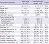
In total, 60 patients were recruited for the study; five were excluded based on the exclusion criteria (Fig. 1). Demographic data and perioperative outcomes are shown in Table 1. No patient complained of any vision change or eye discomfort in the recovery room.
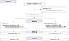
Table 1
Demographic data and perioperative outcomes
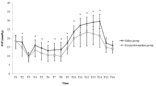
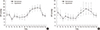
There was no significant difference in the preoperative baseline IOP between the saline and dexmedetomidine groups. The IOP increased sharply after adopting the steep Trendelenburg position, and an increased IOP was maintained during the sustained Trendelenburg position. The IOP at T14 was about 11.3 mmHg higher than that at T1 in the saline group, but only about 4.2 mmHg higher than at T1 in the dexmedetomidine group (Fig. 2A).
Fig. 2
Comparison of intraocular pressure (A) and ocular perfusion pressure (B) between groups. T1 = before anesthetic induction; T2 = before administration of the study drug; T3 = after administration of anesthetic induction agents; T4 = immediately after tracheal intubation; T5-T8 = 1, 3, 5, and 10 minutes after tracheal intubation; T9 = immediately after intraperitoneal CO2 insufflation; T10 = immediately after steep Trendelenburg position; T11-T13 = 1, 2, and 4 h after onset of steep Trendelenburg position; T14 = just before supine position; T15, T16 = 10 and 30 minutes after supine position.
IOP = intraocular pressure, OPP = ocular perfusion pressure.
*Bonferroni correction for multiple comparisons, adjusted P value for significance P < 0.003.
The OPP was reduced during the steep Trendelenburg position in both the saline and dexmedetomidine groups. The degree of decrease in the OPP during the steep Trendelenburg position compared with the baseline OPP was less in the dexmedetomidine group than in the saline group; however, the difference was not statistically significant (18.3 vs. 20.6 mmHg at T14 in the dexmedetomidine and saline groups, respectively; Fig. 2B).
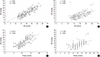
The number of patients with hypertension in the saline group was 13 and that in the dexmedetomidine group was 15. Demographic data between normotensive and hypertensive patients in each group did not show any statistical differences. Patients with underlying hypertension showed slightly higher IOP during the entire period of surgery than normotensive patients in both groups. The difference was not statistically significant (Fig. 3).
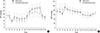
Fig. 3
Comparisons of intraocular pressure between normotensive and hypertensive patients in saline (A) and dexmedetomidine (B) groups. Time values indicated by T1 through T16 are as in Fig. 2.
IOP = intraocular pressure.
The PIP values correlated with the IOP during the surgery (r = 0.881 and 0.739 for the saline and dexmedetomidine groups, respectively; P < 0.001). The Pmean values showed a similar pattern (r = 0.812 and 0.739, respectively; P < 0.001; Fig. 4).
Fig. 4
(A and B) Correlations between peak inspiratory pressure and intraocular pressure. r = 0.881 and 0.739, respectively, for saline (A) and dexmedetomidine groups (B); P < 0.001. (C and D) Correlations between mean inspiratory pressure and intraocular pressure. r = 0.812 and 0.739, respectively, for saline (C) and dexmedetomidine (D) groups; P < 0.001.
DISCUSSION
During laparoscopic lower abdominal procedures in today’s surgical environment, the surgeon’s visualization is assisted by displacing the bowel cephalad away from the surgical field. This can be done by placing the patient in a steep Trendelenburg position. However, an increase in IOP is inevitable in the steep Trendelenburg position. The increased IOP is caused by the increased episcleral venous pressure due to gravity (210).
This study showed that the IOP increased immediately after entering the steep Trendelenburg position, and was not reduced during the sustained position. This differs from the report of Hayreh, who postulated that the IOP would eventually be normalized because of increased drainage of the aqueous humor and decreased episcleral venous pressure that would occur as a result of autoregulatory mechanisms during the steep Trendelenburg position (11). The results of the present study show that it is essential to devise a means to actively attenuate IOP to prevent ophthalmologic complications when it increases significantly during a sustained steep Trendelenburg position. The study results demonstrated attenuation of the increase in IOP with continuous infusion of dexmedetomidine in patients receiving surgery under a steep Trendelenburg position of more than 30°. This effect persisted during the sustained steep Trendelenburg position, indicating that dexmedetomidine is effective in attenuating the increase of IOP associated with this surgical position.
Among various agents known to mitigate IOP, dexmedetomidine, an α-2 agonist, is theoretically considered to be the most appropriate agent for attenuation of the increase in IOP during the steep Trendelenburg position. This is because an α-2 agonist decreases the production of aqueous humor by provoking direct vasoconstriction of afferent vessels in the ciliary body and facilitates the drainage of aqueous humor by decreasing the sympathetically mediated vasomotor tone in the ocular drainage system; the mechanism of the increase in IOP in the steep Trendelenburg position is increased aqueous humor production and episcleral congestion (1213). Additionally, systemically administered α-2 agonists demonstrate a neuroprotective effect on retinal ganglion cell against the increase in IOP. Thus, an α-2 agonist is an appropriate agent for attenuating the increase in IOP and protecting vision (14).
We chose to administer dexmedetomidine continuously rather than as a bolus. Dexmedetomidine has terminal half-life of 2 hours and a relatively shorter context-sensitive half-life, compared with the long surgery duration. Considering this, it should be effective to administer dexmedetomidine continuously during a sustained steep Trendelenburg position. Continuously infused dexmedetomidine in this study contributed to attenuating the increase in IOP during the sustained Trendelenburg position, regardless of the duration of surgery.
Various perioperative factors must be considered that influence IOP in the steep Trendelenburg position. Hemodynamic maintenance, ventilation strategy, and fluid management are factors that are manageable by the anesthesiologist. However, underlying systemic hypertension, body position, CO2 insufflation, and duration of surgery in the steep Trendelenburg position are largely non-adjustable. In this study, we attempted to maintain an intraoperative MAP between ± 20% of the baseline value by modulating the depth of anesthesia, and keep the EtCO2 level between 30 and 35 mmHg by regulating the respiratory rate. Intraoperative fluid was administered according to strict guidelines. By strictly adjusting factors that are manageable by the anesthesiologist, we tried to minimize the influence of those factors on the IOP. As a result, it was possible to evaluate the unadulterated effect of body position, study drugs, and systemic hypertension on the IOP.
Previous studies have not revealed a clear consensus about the relationship between systemic hypertension and the increase in IOP (4151617). We hypothesized that the IOP would be related to the presence of hypertension because ocular perfusion is influenced by vascular dysregulation and abnormal blood pressure (18). However, this study showed no relationship between the IOP and systemic hypertension, consistent with the study of Czarnik et al. (19). Mitchell et al. reported that poorly controlled hypertension increased the risk of open-angle glaucoma, whereas controlled hypertension was not a risk factor for glaucoma (16). In light of this, we attribute our findings to the fact that all patients with underlying hypertension involved in this study were already managed with antihypertensive agents. Furthermore, intravenous agents used for anesthetic induction and continuously administered inhalational anesthetics for anesthetic maintenance played a role in decreasing the IOP, which counteracted the increase in IOP caused by hypertension (20).
Postoperative ophthalmological complications are intimately related to the surgical position (21). Against general expectations, most of these episodes do not appear to be related to direct pressure on the periorbital area or the globe itself, but rather to alterations in the blood flow to the eyeball or the optic nerve, by decreased perfusion or embolism (22). OPP is a commonly used variable to predict the perfusion to the eyeball or optic nerve, which is defined as the difference between the MAP and the IOP. Because the increase in IOP can lower the OPP despite maintenance of a normal MAP, it is important to understand how the IOP and OPP change in anesthetized patients in a steep Trendelenburg position. This study showed that the OPP decreased during the steep Trendelenburg position in both the dexmedetomidine and saline groups, consistent with the results of Molloy (2). However, there was no significant difference in the OPP between the dexmedetomidine and saline groups. This result conflicts with our hypothesis that dexmedetomidine would significantly decrease the MAP and result in a negative influence on the OPP by its sympatholytic effect as an α-2 agonist. The reason why the OPP was maintained despite administration of dexmedetomidine is interpreted in terms of the MAP being maintained in a constant range, especially in hypertensive patients, by regulating the depth of anesthesia diligently with sevoflurane and remifentanil. Patients in present study, especially hypertensive patients, had blood pressure in well-controlled manner. However, there are patients with poorly controlled blood pressure in daily life, and blood pressure tends to be poorly controlled during anesthesia and surgery in those patients. For those patients, maintaining IOP is important to maintain OPP. The anesthesiologist should keep in mind that maintaining a proper MAP by regulating fluid management and the depth of anesthesia and attenuating the increase in the IOP during the steep Trendelenburg position is important for maintaining the OPP and consequently preventing postoperative ophthalmological complications.
We also evaluated the relationships between the PIP or Pmean and the IOP, and found that both the PIP and Pmean were correlated significantly with the IOP. This might be a result of a decreased outflow of aqueous humor through the episcleral vein as a consequence of the increased intrathoracic pressure due to the increase in the PIP. The increased CVP during the steep Trendelenburg position in our study supports this explanation.
We used a hand-held Tono-Pen AVIA tonometer to measure intraoperative IOP. The mechanism of the tonometer is as follows. The tonometer operates on the principle of the Imbert-Fick law: P = F/A, where P = intraocular pressure, F = the amount of force exerted by the tonometer to flatten a specific area of the eye, and A = the area flattened. The tonometer contains a strain gauge that converts IOP measurements to an electrical signal (23). The tonometer was selected as our instrument for measurements because of its speed, ability for use on multiple patients because of its disposable latex tip covers, ease of use, accuracy in a variety of positions, and reliability (24). Setogawa and Kawai (25) measured and compared IOP with an intraocular needle transducer and with a hand-held tonometer in rabbits, and showed a good correlation. Thus, the IOP measured with the Tono-Pen AVIA in this study is trustworthy.
This study has a couple of limitations. Although dexmedetomidine alleviated the increase in IOP in the steep Trendelenburg position, we could not find any objective evidence for decreased incidence or severity of ophthalmological complications due to the use of dexmedetomidine. In fact, no patient complained of postoperative ophthalmological complications despite the increased IOP observed during the steep Trendelenburg position in our study. This might be a result of the relative rarity of ophthalmological complications. According to a literature review, ophthalmological complications, such as visual loss, may occur as a result of sustained increase in IOP during surgery (2627). Thus, we should not overlook the potential harm of a sustained increase in IOP due to the steep Trendelenburg position. Second, we included several kinds of surgeries that may demand different degrees of Trendelenburg position unlike the study of Kim et al. which included the patients undergoing robot-assisted laparoscopic radical prostatectomy (28). However, general, gynecological, and urological surgeons in our institution demand similar degrees of steep Trendelenburg position (30°-45°) for laparoscopic or robot-assisted surgery due to recto-sigmoid colon cancer, prostate cancer, or gynecological cancer. Thus, the effect of different degree of Trendelenburg position might have been minimized.
In conclusion, steep Trendelenburg position during resulted in a considerable increase in IOP and a decrease in OPP during surgery. Continuous infusion of dexmedetomidine is a valuable means for attenuating the increase in IOP without triggering any additional decrease in OPP during surgery, even in sustained steep Trendelenburg position. The IOP in patients with underlying hypertension did not show any significant difference from that in normotensive patients.
 XML Download