

 PDF
PDF ePub
ePub Citation
 Print
Print


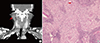
A 49-year-old female presented to our department with right infra-auricular swelling present for more than 3 weeks. The patient’s medical history was not remarkable. The patient had no fever, cough, stridor, dyspnea, shortness of breath, rash, and dryness of mouth or eyes, weight loss, convulsions, or previous hospital admissions. The family history was unremarkable. Physical examination revealed a solitary, firm, non-tender, mobile, and approximately 2-cm sized mass of the right parotid gland. The overlying skin was normal in appearance without erythema or induration. The patient had no cranial nerve deficits and no cervical lymphadenopathy. Computed tomography (CT) scan of the neck demonstrated about 2.2 × 2.1 × 2.2 cm heterogeneously enhancing mass in the right parotid gland (Fig. 1A). Fine-needle aspiration cytology (FNAC) under ultrasound showed chronic granulomatous inflammation. Based on these observations, the preoperative diagnosis was a right-side parotid tumor.
We performed the superficial parotidectomy with preserving facial nerve through a standard ‘lazy S’ cervico-mastoid preauricular surgical incision. The post-operative course was uneventful. The histopathological analysis of the parotid mass demonstrated non-necrotizing granulomatous inflammation suggestive of sarcoidosis (Fig. 1B). Staining of the excised tissues was negative for acid-fast bacilli, and special stains for mycobacteria and fungus revealed no microorganisms. We consulted a pulmonologist and ophthalmologist. Ophthalmologic examinations and radiologic examinations (chest and abdomen CT) revealed no evidence of lung sarcoidosis. The final diagnosis made was parotid gland sarcoidosis. The patient has been regularly followed up usually every 6 months with ophthalmologic and radiologic examinations to check for occurrence of sarcoidosis.
 XML Download