Journal List > J Korean Med Sci > v.31(2) > 1023204

Park, Kwon, Choe, Choi, Oh, Lee, Chong, Kim, Song, Bang, Kim, Kim, Park, Kim, and Oh: Isolation of Middle East Respiratory Syndrome Coronavirus from a Patient of the 2015 Korean Outbreak

Abstract

During the 2015 outbreak of Middle East respiratory syndrome coronavirus (MERS-CoV) in Korea, 186 persons were infected, resulting in 38 fatalities. We isolated MERS-CoV from the oropharyngeal sample obtained from a patient of the outbreak. Cytopathic effects showing detachment and rounding of cells were observed in Vero cell cultures 3 days after inoculation of the sample. Spherical virus particles were observed by transmission electron microscopy. Full-length genome sequence of the virus isolate was obtained and phylogenetic analyses showed that it clustered with clade B of MERS-CoV.

Middle East respiratory syndrome coronavirus (MERS-CoV) is a betacoronavirus causing a severe acute respiratory infection (1,2). It was first isolated from the sputum of a patient with severe pneumonia in Saudi Arabia in 2012 (3). Since then, 26 countries have reported 1,618 laboratory-confirmed cases of infection with MERS-CoV to the World Health Organization (WHO), including 579 fatalities (4).
The Korean outbreak of MERS-CoV was initiated in May 2015 by a business man returning from the Middle East (5). The transmission of MERS-CoV continued until early July, resulting in 186 cases with 38 deaths. One of the most important characteristics of the Korean outbreak was 4 large clusters of cases due to superspreading event at hospitals, accounting for > 80% of the total cases. Another characteristic was that many cases of second- and third-generation of transmission occurred (5,6). This finding is quite contrast to the previous studies suggesting limited person-to-person transmissibility of MERS-CoV (7,8). To better understand transmissibility and assess epidemic risk, characterization of MERS-CoV of the Korean outbreak would be of paramount importance (9). Here, we report the MERS-CoV isolated from a patient of the Korean outbreak.
A 39-year-old healthcare worker was admitted to the hospital because of fever and cough. On May 27, 2015, he was unknowingly exposed to the index case (designated as patient number 14 by Korea Ministry of Health and Welfare) of the hospital outbreak of Middle East respiratory syndrome coronavirus (MERS-CoV) at emergency department of a hospital (10). Two days later, he developed fever and dry cough. On June 2, he was diagnosed with MERS-CoV infection as sputum sample was positive on real-time reverse transcriptase polymerase chain reaction (RT-PCR) assay, and admitted to the isolation unit of the MERS-designated hospital by the government.
He had a history of cough variant asthma, but did not take any regular medication, and otherwise healthy. On admission (June 2, 2015), the physical examination revealed a body temperature of 38.8°C, a respiratory rate of 22 breaths per minute, a pulse of 78 per minute, and a blood pressure of 118/71 mmHg. Chest radiography showed patchy consolidation in the upper zone of the left lung. His pneumonia progressed, and on June 8, he developed shortness of breath, his arterial oxygen saturation decreased below 90%, requiring oxygen supplementation, and chest radiography showed multiple consolidations in the both lungs. On June 10, he was intubated and mechanical ventilation was started. His hypoxemia worsened rapidly, and veno-venous extracorporeal membrane oxygenation support was started since June 11. On July 2 (day 35 of his illness), real-time RT-PCR for MERS-CoV turned negative, and was removed from the isolation unit. He recovered gradually.
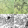
The patient’s oropharyngeal samples were obtained by using UTMTM kit containing 3 mL of viral transport media (Copan Diagnostics Inc., Murrieta, CA). The samples were stored at -70°C until assays. We inoculated monolayers of Vero cells with the samples and cultured the cells at 37°C in a 5% carbon dioxide atmosphere. Cytopathic effects consisting of rounding and detachment of cells were observed 3 days after the inoculation of the sample taken on day 11 of his illness (Fig. 1A and B). The RNA titer in the sample was 5.80 × 10⁷ copies/mL for upE gene and 4.97 × 10⁷ copies/mL for ORF1a gene.
In order to observe virus particles, Vero cell monolayer showing the cytopathic effects was fixed as previously described (11). It was cut on ultramicrotome (RMC MT-XL) at 65 nm. Ultrathin sections were stained with saturated 4% uranyl acetate and 4% lead citrate before examination with a transmission electron microscope (JEM-1400; JEOL USA Inc., Peabody, MA, USA) at 60 kV. Spherical particles ranging 77 to 131 nm in diameter were observed within the cytoplasm of infected cells (Fig. 1C and D).
For full-length genome sequencing of the virus isolate (MERS-CoV Hu/KOR/SNU1_035/2015), Vero cell monolayer showing cytopathic effects was harvested and used for RNA extraction. RNA was extracted by using QIAamp viral RNA mini kit (QIAGEN, Valencia, CA), according to the manufacturer’s instructions. The RNA was used for cDNA synthesis using SuperScript III Reverse Transcriptase (Invitrogen, MA, USA) by each specific RT primer as described previously (12). Finally, about 2.5 kb PCR products were amplified by each primer pair (Table 1), and the amplicons were sheared by Covaris S2 according to the 200 bp target BP condition (Covaris, MA, USA). To generate the Next Generation Sequencing (NGS) library, the fragments were ligated with adapter and index (barcode) using TruSeq Nano DNA HT Library Prep kit (Illumina, CA, USA), and the library was sequenced by MiSeq (Illumina, CA, USA). The NGS data were aligned to MERS-CoV, NC_019843, used for Binary Sequence Alignment/Map (BAM) file generation, and genome assembly. In order to evaluate genetic relationship between this isolate and Homo sapiens and Camelus dromedaries MERS-CoV sequences reported from other countries, phylogenetic analyses were conducted using the whole genome, the S gene and the OFR1a gene.
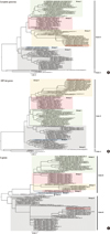
The full-length genome sequence (30,119 bp) of the virus isolate was obtained and deposited in the GenBank (accession no. KU308549). The genome sequence of the virus had high level of nucleotide identity (97.80%–99.95%) to those of MERS-CoV reported previously (Fig. 2A). Of note, the closest ones were KOREA/Seoul/035-1-2015 and 035-2-2015 (GenBank accession no. KT374054–5), that were directly sequenced from sputum of the same patient as ours (13). A previous study about S gene of MERS-CoV reported from Korea showed that a culture isolate from patient number 002 contained two nonsynonymous variants (S137R and V530L) (14). These variants were not found in our isolate and there was no difference in amino acids of S protein between our isolate and directly sequenced ones (KT374054-5). This difference can be explained by cell culture-adaptation in that our culture isolate was obtained before passage whereas one with nonsysnonymous variant was from the third passage in Vero cells.
Phylogenetic analyses of the whole genome showed that this virus closely clustered with those reported from Korea (GenBank accession nos. KT029139, KT374050– KT374057), China (GenBank accession no. KT006149.2) and Saudi Arabia in 2015 (GenBank accession no. KT026453–6). Phylogenetic analyses based on ORF1ab genes revealed that this virus fell into the group 3, but those based on S genes showed that this virus belongs to the group 5 along with other viruses reported from Korea (Fig. 2B and C). These findings are compatible to a previous study (15).
In summary, we isolated MERS-CoV from a patient with severe pneumonia who had been infected during the Korean outbreak in 2015. We also obtained full-length sequence of the virus isolate. Phylogenetic analyses showed that the isolate belongs to clade B of MERS-CoV.

Figures and Tables

Fig. 1

Cytopathic effects of MERS-CoV in Vero cell cultures and Electron microscopy image of MERS-CoV. Vero cells were inoculated with oropharyngeal swab sample. (A) Vero cell cultures in negative control. (B) Cytopathic effects (rounding and detachment of cells) in Vero cell cultures 3 days after inoculation of the sample. (C, D) Transmission electron microscopy image of Vero cells infected with MERS-CoV. White arrow denotes nuclear membrane. Brown scale bar indicates 100 μm (A and B). Black scale bar indicates 500 nm (C) and white scale bar does 200 nm (D).
jkms-31-315-g001
Fig. 2

Molecular phylogenetic analysis. Phylogenetic tree on complete genome (A), S genes (B), and ORF1ab genes (C) for the 101 gene sequences of MERS-CoV. The evolutionary history was inferred by using the maximum likelihood method based on the Tamura-Nei model (16). Evolutionary analyses were conducted in MEGA6 (17). Red box indicates our virus isolate.
jkms-31-315-g002
Table 1

PCR primer pairs used in this study

jkms-31-315-i001
No. Forward sequence (5'-->3') Reverse sequence (5'-->3')
1 FEP_1_F GATTTAAGTGAATAGCTTGGCTATCTC FCO_TM58_70342_1_R GGAATATTAGAGACTCCCTGCCG
2 2497F TCCCATCGGGAACCTATTACTGTG FCO_TM58_68466_R TGTAACCACCATTAGTGCGGAC
3 4477F ACGTTAAGTTAAACCCTTCAGAAG FCO_TM58_67825_1_R AATGAAGCCCTAAATAGTAACTTCACT
4 new2_15F_6427 TGCTTAGATTGCACACCGTT FCO_TM58_68526_R AGGTGGTTAACCGGAAAGCTAAA
6506F AGAATTTGCTACCCGCACTTTCACTG FCO_TM58_68526_R AGGTGGTTAACCGGAAAGCTAAA
5 new2_19F_8393 GGATGCACTTAAACGACAGA FCO_TM58_33984_R GTGTAACCAACACTACCACAAGAAC
8496F TGGTGCTCCTACATGGTTTAATGCG FCO_TM58_33984_R GTGTAACCAACACTACCACAAGAAC
6 10477F ACACCAAGGAGGGTAGTGTGATC FCO_TM58_67887_R GAATTACAACGCGAAGTTTATTTGAAG
7 FCO_TM58_2977_F AAGGCTTTGCAGAAGGCTGTTA FCO_TM58_68115_R GAATTACAACGCGAAGTTTATTTGAAG
12488F AGGTAGTCACATATCCCTCGCTTAAC FCO_TM58_68115_R TCATCAACTCCTTAAGAGAGAGCCTAT
8 14481F TGGATGTTAGTCTCCATAGACATAG FCO_TM58_34102_R TGTAATCACCACTTTCAGTCCAGT
9 16476F TCGGCTTATACAAGAATATGTGCAC FCO_TM58_981_R CATGAGCCCAACAAACAAACGTA
10 18490F ACGACGTATAGTGCAAATGTTGTC FCO_TM58_69145_R AGCTTTAAATCTATAACAGAACACACC
11 new2_42F_20357 AAGAAGCAACAGGAAGGTCA FCO_TM58_34878_R TAGAAGGCAGCCCAAGCTTTT
20490F AAGACCTTGGCGTAGTATCCAAGG FCO_TM58_34878_R TAGAAGGCAGCCCAAGCTTTT
12 FCO_TM58_67088_1_F CCACCTTGCCTGTTTATGATACTATTA FCO_TM58_72579_R CTGTTTGCATAGCTCCCAGAG
22481F TGATTTGTCACAACTCCACTGC FCO_TM58_72579_R CTGTTTGCATAGCTCCCAGAG
13 FCO_TM58_66781_F TGGACTGCTGGCTTATCCTC FCO_TM58_66820_R GCTTAAATCTATGTATGTTAGCACAGT
24512F TCAGAAGGTTCAGGATGCTGTGAAC FCO_TM58_66820_R GCTTAAATCTATGTATGTTAGCACAGT
14 26470F TGAGTTCGCCTTGCTGCGCAAAAC FCO_TM58_69858_R TGTAATTACCTGCCTTATATCTATGGT
15 new2_59F_28427 GGCAAAGCTACGGAACTAAT FEP_3_R GCAAATCATCTAATTAGCCTAATCTAATTG
28490F AACTTGCATTGCTTCGAGCTTAGG FEP_3_R GCAAATCATCTAATTAGCCTAATCTAATTG

ACKNOWLEDGMENT

We thank Ms. Myoung Im Shin (Department of Pathology, Seoul National University Hospital) for her technical assistance in electron microscopy.

Notes

Funding This work was supported by a grant from the Korean Healthcare Technology R&D Project through the Korea Health Industry Development Institute (KHIDI), funded by the Ministry of Health & Welfare, Republic of Korea (grant number: HI15C3227).

DISCLOSURE The authors have no potential conflicts of interest to disclose.

AUTHOR CONTRIBUTION Conception and design: Oh MD, Park WB. Acquisition of data: Park WB, Kwon NJ, Choe PG, Oh HS, Choi SJ, Lee SM. Analysis and interpretation of data: Oh MD, Park WB, Chong H, Kim JI, Song KH, Bang JH, Kim ES, Kim HB, Park SW, Kim NJ. Manuscript preparation: Oh MD, Park WB, Kwon NJ, Choe PG. Manuscript approval: all authors.

References

1. Chan JF, Lau SK, To KK, Cheng VC, Woo PC, Yuen KY. Middle East respiratory syndrome coronavirus: another zoonotic betacoronavirus causing SARS-like disease. Clin Microbiol Rev. 2015; 28:465–522.
2. Zumla A, Hui DS, Perlman S. Middle East respiratory syndrome. Lancet. 2015; 386:995–1007.
3. Zaki AM, van Boheemen S, Bestebroer TM, Osterhaus AD, Fouchier RA. Isolation of a novel coronavirus from a man with pneumonia in Saudi Arabia. N Engl J Med. 2012; 367:1814–1820.
4. World Health Organization. Middle East respiratory syndrome coronavirus (MERS-CoV) - Saudi Arabia: disease outbreak news. updated on 4 December 2015. Available at http://www.who.int/csr/don/4-december-2015-mers-saudi-arabia/en/.
5. Korea Centers for Disease Control and Prevention. Middle East respiratory syndrome coronavirus outbreak in the Republic of Korea, 2015. Osong Public Health Res Perspect. 2015; 6:269–278.
6. Chowell G, Abdirizak F, Lee S, Lee J, Jung E, Nishiura H, Viboud C. Transmission characteristics of MERS and SARS in the healthcare setting: a comparative study. BMC Med. 2015; 13:210.
7. Breban R, Riou J, Fontanet A. Interhuman transmissibility of Middle East respiratory syndrome coronavirus: estimation of pandemic risk. Lancet. 2013; 382:694–699.
8. Cotten M, Watson SJ, Kellam P, Al-Rabeeah AA, Makhdoom HQ, Assiri A, Al-Tawfiq JA, Alhakeem RF, Madani H, AlRabiah FA, et al. Transmission and evolution of the Middle East respiratory syndrome coronavirus in Saudi Arabia: a descriptive genomic study. Lancet. 2013; 382:1993–2002.
9. Edelstein M, Heymann DL. What needs to be done to control the spread of Middle East respiratory syndrome coronavirus? Future Virol. 2015; 10:497–505.
10. Oh MD, Choe PG, Oh HS, Park WB, Lee SM, Park J, Lee SK, Song JS, Kim NJ. Middle East respiratory syndrome coronavirus superspreading Event Involving 81 Persons, Korea 2015. J Korean Med Sci. 2015; 30:1701–1705.
11. Graham L, Orenstein JM. Processing tissue and cells for transmission electron microscopy in diagnostic pathology and research. Nat Protoc. 2007; 2:2439–2450.
12. Cotten M, Lam TT, Watson SJ, Palser AL, Petrova V, Grant P, Pybus OG, Rambaut A, Guan Y, Pillay D, et al. Full-genome deep sequencing and phylogenetic analysis of novel human betacoronavirus. Emerg Infect Dis. 2013; 19:736–742B.
13. Seong MW, Kim SY, Corman VM, Kim TS, Cho SI, Kim MJ, Lee SJ, Lee JS, Seo SH, Ahn JS, et al. Microevolution of outbreak-associated Middle East respiratory syndrome coronavirus, South Korea, 2015. Emerg Infect Dis. Forthcoming. 2016.
14. Kim DW, Kim YJ, Park SH, Yun MR, Yang JS, Kang HJ, Han YW, Lee HS, Man Kim H, Kim H, et al. Variations in spike glycoprotein gene of MERS-CoV, South Korea, 2015. Emerg Infect Dis. 2016; 22:100–104.
15. Wang Y, Liu D, Shi W, Lu R, Wang W, Zhao Y, Deng Y, Zhou W, Ren H, Wu J, et al. Origin and possible genetic recombination of the Middle East respiratory syndrome coronavirus from the first imported case in China: phylogenetics and coalescence analysis. MBio. 2015; 6:e01280–15.
16. Tamura K, Nei M. Estimation of the number of nucleotide substitutions in the control region of mitochondrial DNA in humans and chimpanzees. Mol Biol Evol. 1993; 10:512–526.
17. Tamura K, Stecher G, Peterson D, Filipski A, Kumar S. MEGA6: Molecular evolutionary genetics analysis version 6.0. Mol Biol Evol. 2013; 30:2725–2729.
TOOLS
ORCID iDs

Wan Beom Park
https://orcid.org/http://orcid.org/0000-0003-0022-9625

Nak-Jung Kwon
https://orcid.org/http://orcid.org/0000-0003-0874-4146

Pyoeng Gyun Choe
https://orcid.org/http://orcid.org/0000-0001-6794-7918

Su-Jin Choi
https://orcid.org/http://orcid.org/0000-0001-8732-3950

Hong Sang Oh
https://orcid.org/http://orcid.org/0000-0002-4535-6305

Sang Min Lee
https://orcid.org/http://orcid.org/0000-0002-1388-9318

Hyonyong Chong
https://orcid.org/http://orcid.org/0000-0001-6928-8805

Jong-Il Kim
https://orcid.org/http://orcid.org/0000-0002-7240-3744

Kyoung-Ho Song
https://orcid.org/http://orcid.org/0000-0002-4517-3840

Ji Hwan Bang
https://orcid.org/http://orcid.org/0000-0002-7628-1182

Eu Suk Kim
https://orcid.org/http://orcid.org/0000-0001-7132-0157

Hong-Bin Kim
https://orcid.org/http://orcid.org/0000-0001-6262-372X

Sang Won Park
https://orcid.org/http://orcid.org/0000-0002-0550-1897

Nam Joong Kim
https://orcid.org/http://orcid.org/0000-0001-6793-9467

Myoung-don Oh
https://orcid.org/http://orcid.org/0000-0002-2344-7695

Similar articles