

 PDF
PDF ePub
ePub Citation
 Print
Print


INTRODUCTION
Acute eosinophilic pneumonia (AEP), first described by Allen et al. in 1989 as a distinct disease entity, is an uncommon inflammatory lung disease (1). AEP presents with acute-onset respiratory symptoms, diffuse radiographic infiltrates, and eosinophilic infiltration into the lungs without a known cause of pulmonary eosinophilia, such as drugs, toxins, or parasite infestations (234). The clinical severity of respiratory distress associated with AEP varies from mild to severe; however, most patients with AEP exhibit rapid responses to systemic corticosteroid therapy without sequelae, even in fatal cases (5678).
Recent studies concerning the clinical features of AEP have suggested that unidentified inhaled antigens could be partly responsible for the inflammatory process (91011121314), and recent molecular studies have revealed that several inflammatory cytokines are associated with eosinophil recruitment into the lungs, degranulation, and eosinophil survival (1516171819). However, to date, limited data exist concerning the detailed clinical features of AEP and possible factors in its occurrence, limiting insights into its epidemiology or pathophysiology.
In Korean military hospitals, several cases of AEP are diagnosed and managed annually, and an abundance of medical records and demographic data regarding the characteristics and outcomes of AEP patients has accumulated (562021). Thus, in this study, we retrospectively investigated the clinicodemographic data of AEP cases managed in Korean military hospitals to evaluate clinical characteristics and associated factors that contribute to the development of, or increased vulnerability to, AEP.
MATERIALS AND METHODS
Study population
We retrospectively reviewed the medical records of consecutive adult patients with newly diagnosed AEP between January 2007 and December 2013 at multiple Korean military hospitals, including The Armed Forces Capital Hospital, The Armed Forces Busan Hospital, The Armed Forces Cheongpyeong Hospital, The Armed Forces Chuncheon Hospital, The Armed Forces Daegu Hospital, The Armed Forces Daejeon Hospital, The Armed Forces Gangneung Hospital, The Armed Forces Goyang Hospital, The Armed Forces Hampyeong Hospital, The Armed Forces Hongcheon Hospital, The Armed Forces Ildong Hospital, The Armed Forces Seoul Hospital, The Armed Forces Wonju Hospital, The Armed Forces YangJu Hospital, The Naval Marine Medical Center, The Naval Pohang Hospital, and The Air Forces Aerospace Medical Center.
A definitive diagnosis of AEP was based on a modification of the criteria proposed by Philit et al. (2), as reported previously (5): 1) acute onset of febrile respiratory manifestations <1 month in duration; 2) bilateral diffuse infiltrates on chest radiography; 3) >25% eosinophils in bronchoalveolar lavage (BAL) or eosinophilic pneumonia on lung biopsy; and 4) absence of known causes of pulmonary eosinophilia, including drugs, toxins and infections. If bronchoscopy with BAL or lung biopsy was not available, a clinical diagnosis of AEP was made when a rapid response to systemic corticosteroid therapy was achieved, in addition to definitive diagnostic criteria 1), 2), and 4). All patients with a definitive and clinical diagnosis of AEP were included in the analysis.
Patient management and data collection
Patients who were clinically suspected of AEP underwent a diagnostic workup consisting of laboratory examinations, chest radiography, computed tomography (CT), and/or flexible bronchoscopy with BAL. Infectious etiologies were investigated using peripheral blood, sputum, tracheal aspirates and BAL fluid using the following techniques: staining and microbiological culture for bacteria and Mycobacterium tuberculosis; multiplex polymerase chain reaction for respiratory viruses, including influenza virus, parainfluenza virus, adenovirus and respiratory syncytial virus; and serological testing for atypical pathogens, including Mycoplasma pneumoniae, Chlamydia pneumoniae, and parasites using specific antibody tests. Whether diagnostic tests or interventions were performed was decided by the attending physician.
Most patients diagnosed with AEP were treated with systemic corticosteroid. Before May 2007, the dose or duration of corticosteroid tapering was not standardized, while after May 2007, 2- or 4-week tapering protocols were widely used (5). After the publication of a study in May 2012 showing the non-inferiority of 2-week corticosteroid treatments compared to 4-week corticosteroid treatments in managing AEP patients (5), 2-week tapering protocols became widely applied to AEP patients. An example of the 2-week corticosteroid treatment protocol is as follows: the initial dose of corticosteroid is chosen based on the presence of respiratory failure, defined as a partial pressure of arterial oxygen (PaO2)/fraction of inspired oxygen (FiO2) ratio ≤300 and/or tachypnea (respiration rate >30 breaths/min). Patients with respiratory failure received 60 mg methylprednisolone intravenously every 6 hours for 3 days followed by 30 mg oral prednisolone twice daily for 4 days. Patients without respiratory failure received 30 mg oral prednisolone twice daily for 7 days, followed by tapering over 2 weeks. However, when oxygenation was not severely impaired (PaO2/FiO2 ratio >350) and symptoms were mild, the patient underwent conservative treatment without corticosteroids based on the decision of the attending physician.
The following data were collected retrospectively: demographic characteristics including age, sex, body mass index, underlying diseases, history of recent upper-respiratory infections, smoking habits, and working position in military services. Clinico-radiological findings and laboratory data at initial presentation were collected, and clinical parameters that reflect disease severity, including oxygen requirement rate, intensive care unit (ICU) admission rate, the need for mechanical ventilation, and pneumonia severity index score, were also collected. Annual incidence rates and seasonal variations were also evaluated.
In calculating annual incidence rates, we used the cited population of each military service as a denominator, which is published biennially by The Ministry of National Defense, Korea, in the defense white paper. This was used to calculate annual incidence rates of AEP among the entire military population and within each service. A Poisson-distribution was applied to estimate confidence intervals since most medical events are considered to follow Poisson-distributions, especially rare diseases like AEP. Follow-up data were last obtained on February 1, 2014. All medical records were anonymized or de-identified.
Statistical analysis
Data are presented as medians and interquartile ranges (IQR) for continuous variables and as numbers and percentages for categorical variables. Data were compared using the Kruskal-Wallis test (more than two independent groups) for continuous variables, and a χ2 or Fisher's exact test for categorical variables. Values of P<0.05 were considered to indicate statistical significance. All statistical analyses were performed using the PASW software (ver. 18.0; SPSS Inc., Chicago, IL, USA).
Ethics statement
The study protocol was designed in accordance with the Declaration of Helsinki and was approved by the institutional review board of the Armed Forces Capital Hospital (AFMC-14-IRB-016) and the Armed Forces Medical Command on behalf of the military hospitals that were involved in the current study. Informed consent was exempted by the board.
RESULTS
Clinical characteristics of patients with AEP
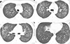
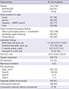
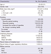
Three hundred and thirty-three patients with AEP were identified in the study period, including 304 (91%) definitive and 29 (9%) clinical AEP patients. Clinical characteristics of study patients are shown in Table 1. All patients had acute respiratory symptoms or signs including cough, sputum, dyspnea, or fever. On initial chest computed tomography scans, all patients had diffuse ground glass opacity with or without consolidation (Fig. 1), 282 (85%) had interlobular septal thickening, and 265 (80%) had pleural effusion. All patients had elevated inflammatory markers, including white blood cell count and C-reactive protein, with median values of 13,185 (IQR, 9,700-17,697)/µL and 9.51 (IQR, 5.72-14.4) mg/dL, respectively, and median peripheral eosinophil counts of 314 (182-538)/µL. The median value of the PaO2/FiO2 ratio was 276.2 (IQR, 239.3-329.0), 32% of the patients had severe hypoxemia (PaO2/FiO2 <250), and 258 (78%) patients had an oxygen requirement. Of the study patients, 103 (31%) were admitted to the ICU, 8 (2%) required mechanical ventilation support, and approximately one-third of the study patients had more than a pneumonia severity index score (PSI) class II; class III (n=86; 26%), class IV (n=4; 1%), and class V (n=1; 1%). Overall, 306 (92%) AEP patients were treated with systemic corticosteroid and 27 (8%) patients did not receive corticosteroid; all patients survived without sequelae.
Demographics of patients with AEP
The demographics of AEP patients are shown in Table 2. The median age was 22 (IQR, 20-21) years and all patients were male. The median body mass index was 22.9 (IQR, 21.2-24.6 kg/m2); 67 (20%) patients were overweight (>25 kg/m2) and 11 (3%) patients were underweight (<18.5 kg/m2). Most patients were current smokers (n=309; 93%); among these, 175 (53%) patients had recently started smoking, 100 (30%) patients had resumed smoking, and only 24 (7%) patients were ex-smokers or had never smoked. Only 30 (9%) patients had underlying diseases; allergic rhinitis (n=17; 5%) was the most common, followed by asthma (n=9; 3%) and atopic dermatitis (n=4; 1%). One patient with a recent history of inhalation burns and one patient with a recent upper respiratory infection were identified. Most of the patients were in the Army (n=297; 89%), with 29 (9%) in the Navy and only 7 (2%) in the Air Force.
Annual incidences of AEP occurrence according to military service
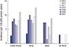
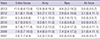
Because absolute numbers of AEP patients were much lower in the Navy and Air Force than in the Army, as shown by the demographics of study patients, we calculated and compared the annual incidence of AEP occurrence according to military service (Table 3). Among all forces, in 2008 the incidence of AEP abruptly increased compared to 2007, and by 2013 the incidence of AEP was highest at up to 11.0 per 100,000 person-years (95% confidence interval, 8.6-13.9; Fig. 2). Interestingly, in each year, the incidence of AEP tended to be higher among Army personnel compared to Navy or Air Force personnel, and these differences were statistically significant (Kruskal-Wallis, P=0.002). However, the differences in other clinico-demographic data between the different military services were not statistically significant (data not shown).
Seasonal variation in the occurrence of AEP patients
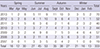
Since physicians working at military hospitals generally estimate that more cases of AEP are encountered in the summer than in other seasons, we investigated seasonal variations in the occurrence of AEP among all military services to estimate whether occurrence was influenced by season (Table 4). Of all AEP cases, 125 (38%) occurred during the summer, 100 (30%) during the autumn, 58 (17%) during the spring, and only 50 (15%) during the winter. Numbers of AEP patients tended to increase as seasonal temperatures increased (Table 4) and peaked in the summer each year, around August.
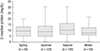
We additionally evaluated the number of AEP patients with a relatively high severity of illness according to season (Table 5). The number of severe cases tended to increase as seasonal temperatures rose and peaked around the summer, from late spring to early autumn. The numbers of patients who needed oxygen (n=99) and ICU admission (n=38), and had a PSI score of III (n=31) or IV (n=3) tended to be higher in summer than in other seasons. None of the patients who needed mechanical ventilation was seen during the winter. One of the most severe cases of AEP (PSI class V) occurred during late spring, in May. However, differences in other clinico-demographic data according to season (spring vs. summer vs. autumn vs. winter) were not statistically significant (data not shown). The initial level of C-reactive protein in AEP patients had a slight tendency to be higher during summer and autumn than during spring and winter (Fig. 3), and median values of C-reactive protein in spring, summer, autumn, and winter were 9.53 (IQR, 5.34-13.1) mg/dL, 9.04 (IQR, 4.90-13.8) mg/dL, 10.10 (IQR, 6.62-16.02) mg/dL, and 7.90 (IQR, 5.96-12.61) mg/dL, respectively; however the differences did not reach statistical significance.
DISCUSSION
In this study, we investigated the clinical characteristics of AEP and evaluated possible demographic factors that may contribute to its development in Korean military personnel. Our data suggest that several environmental factors such as recently changed smoking habits, military service in the Army, and increasing seasonal temperature could be related to the occurrence of AEP. Regarding environmental influences on the development of AEP, previous studies have indicated that inhaled stimulants may trigger the inflammatory processes of AEP (9101112). For example, an observational study that included 33 AEP patients found that altered smoking habits, not only starting to smoke but also restarting smoking and increasing daily smoking doses, were associated with the development of AEP (12). Moreover, Rom et al. presented a case study of a firefighter that developed AEP after exposure to dust from the World Trade Center collapse (10). Similar to these previous studies, the majority of our patients (n=288; 87%) had recently altered smoking habits and the annual incidence of AEP was highest in the Army, which may be because the Army has more ground services, and outdoor activity in mountainous areas of Korea may increase the likelihood of inhaling unidentified antigens.
Interestingly, in this study we found that the occurrence of AEP was related to seasonal variations. The number of AEP patients tended to increase as seasonal temperatures rose and peaked around summer, which was associated with greater disease severity than the other seasons. Moreover, AEP with relatively high illness severity was more common in the summer than in other seasons, although seasonal differences in the parameters that indicate disease severity did not reach statistical significance. These phenomena could be partly explained by the rise in temperature as the season changes from winter to summer, possibly increasing the likelihood of inhalation of stimulants or unidentified antigens, which in turn may trigger lung inflammation. One other study has described an association between AEP occurrence and season or climate. When evaluating the clinical features and epidemiology of 18 AEP cases in the United States military personnel deployed in or near Iraq, summer had the highest incidence of AEP (22). However, limited data exist concerning environmental influences on AEP occurrence, and well-designed studies of its occurrence that adjust for other confounding factors have not been performed, mainly due to the rarity of the disease, which limits insight into its epidemiology or pathophysiology.
In our study only a small proportion (n=11; 3%) of patients had a low body mass index (<18.5 kg/m2) and the majority of patients (n=255; 77%) had a normal body mass index. This body mass index distribution is uncommon in other pulmonary diseases such as tuberculosis and other inflammatory pulmonary diseases. Additionally, only 9% of study patients had underlying allergic diseases. These findings suggest that inflammation in AEP is not a chronic process and that AEP may have different inflammatory processes than known allergic reactions. To date there has not been a comprehensive study of the association of AEP with immunoglobulin E-mediated or allergic reactions. The results of a recent study indicated that immunoglobulin G levels decreased significantly during active disease states and increased during remission, but serum immunoglobulin E levels did not change significantly, suggesting that the pathogenesis of AEP may negatively impact serum immunoglobulin G levels but not immunoglobulin E levels (23). However, accurate data concerning the inflammatory process are limited; thus, further pathophysiological studies along with epidemiological data are needed.
In our study, the incidence of AEP in recent years was similar to previously reported rates. In a study of the characteristics and epidemiology of AEP among the United States military personnel, Shorr et al. (22) reported an AEP incidence of 9.1 per 100,000 person-years (95% confidence interval, 4.3 to 13.3). However, in our current study the annual AEP incidence rate varied from 2.8 per 100,000 person-years in 2007 to 11.0 per 100,000 person-years in 2013. The lower incidence in 2007 compared to other years is likely due to inadequate awareness of AEP in military hospitals in Korea. The peak incidence of AEP in 2013 may be due to improved insight into AEP by military physicians as well as the increased performance of diagnostic flexible bronchoscopies using the BAL procedure in unknown respiratory failure patients. Therefore, more aggressive diagnostic evaluation of patients will facilitate determination of accurate incidence rates, changes in AEP incidence rates, and aid the investigation of its epidemiology.
This study has some limitations. First, there was no matched normal control group to compare to the AEP group since we performed a retrospective case series study. This type of study was chosen since collecting medical data from healthy military personnel is difficult as it is not permitted without definite medical evidence in any of the Korean armed forces. Thus, based on the results of this study, we evaluated only accurate case series data. Second, because our study was conducted at military hospitals only, most patients were previously healthy males with a uniform median age, and thus not representative of the general population. Third, there is a possibility that the incidence or occurrence of AEP might have been underestimated in our study population because some patients might have been treated at a local military hospital or might have improved spontaneously. However, we cautiously estimate that this influence is not significant due to the unique characteristics of the medical system of the Korean military services. According to the military law of Korean military personnel, all soldiers should be treated in military hospitals first if possible. Accordingly, most patients with suspected AEP were likely transferred to a military hospital, including the Armed Forces Capital Hospital, which is the largest tertiary referral military hospital in Korea, where bronchoscopy is available.
In conclusion, in this study we investigated the clinical characteristics of AEP, demonstrated that patients with AEP frequently have recently changed smoking habits and work for the Army, and found an increasing tendency in the numbers of patients and of patients with higher AEP severity with increasing seasonal temperatures. These results highlight the need for future pathophysiological and epidemiological prospective studies to evaluate the impact of external environmental variables on the occurrence and pathophysiology of AEP.

 XML Download