

 PDF
PDF ePub
ePub Citation
 Print
Print


INTRODUCTION
Among various microbial pathogens, Mycobacterium tuberculosis (TB) infection has been regarded as an important subject of scientific studies on the diseases prevalent among pre-modern peoples. By evidences of anatomical pathology, researchers could report the presence of TB in the archaeologically obtained human samples. Using the ancient samples, the phylogeny of TB continues to have been traced by the molecular biological techniques (1234). Although more historical knowledge on TB infection has been obtained by studies on archaeological samples worldwide, there were very few reports on the presence of TB cases from ancient human remains in Korea. Considering that TB was known to have been endemic in this country until quite recent days, the deficiency of medical evidences on the TB infection in archaeological samples of Korea looks exceptional. In this regard, the current case about calcified pulmonary nodules (PN) that were recently identified in 350-yr-old-Korean mummy is very meaningful to concerned researchers. By differential diagnosis based upon CT radiological findings, we can conclude that this looks the first-ever case of pulmonary TB identified among archaeologically obtained, pre-modern Korean samples.
CASE DESCRIPTION
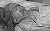
A female mummy was discovered in April 2010, from a Joseon tomb discovered in Mungyeong County of Korea (Fig. 1). By the tree-ring test for the coffin wood, the tomb was estimated to have been constructed in around the 1650s CE. After the mummy was moved to our laboratory, the sex determination was made as a female, by its external genitalia and by morphology of the pelvic bone (5). The mummy's age was estimated to be 40-44 yr old, by the degeneration pattern of the auricular and pubic symphyseal surfaces of the hipbones (6). The age was also estimated by the method of Lamendin et al. (1992) (7) on right maxillary first premolar as about 47.2 yr-old.
Next, the CT scan was performed on this case with a 64 MDCT scanner (VCT, GE Healthcare, Milwaukee, WI, USA) at Seoul National University Hospital, Korea. Using the helical technique (120 kVP), a spiral volume from head to toe was acquired. All of the data were reconstructed into axial images representing 1.25 mm thicknesses at 1.25 mm intervals. These images were transferred to a workstation (Advantage Windows Workstation 4.3; GE Healthcare) preparatory to post-processing, by which coronal and sagittal multi-planar reformation and volume-rendering images were obtained. Volume-rendering images were obtained by InVesalius 3.0.0 Beta 5 (CTI, Campinas, Brazil).
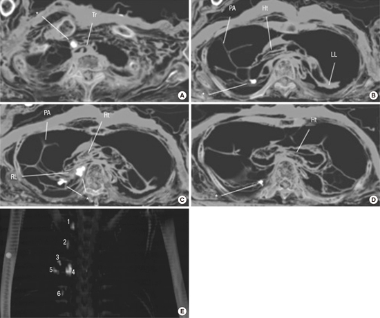
The CT images exhibited six radiopaque nodules in right-sided lung within thoracic cavity, at the level of thoracic vertebrae (TV) 1 to 6 (Fig. 2). Briefly, at TV1 level, a radiopaque nodule (#1) was located in front of trachea, especially at the right side of it (Fig. 2A). A small-sized nodule (#2) was also identified at TV3, along the trachea. Another radiopaque nodule (#3) was found in the mummified lung (TV4 level) that was displaced to posterior thoracic cavity (Fig. 2B). At TV5 level, two large nodules (#4 and #5) were observed around right pulmonary hilum (Fig. 2C). The nodule #6 was found in the right lung, around costovertebral junction at TV6 level (Fig. 2D). Volume rendering images show this calcification pattern clearly in Fig. 2E. From TV1 to TV7 level, we found presumptive pleural adhesions on CT, mainly in the right-sided thoracic cavity.
For post-factum validation of the CT findings, we dissected the mummy in the Department of Anatomy, Seoul National University College of Medicine (Korea). We made an incision along the lower borders of the xiphoid process and the 12th ribs. From the lower tip of the xiphoid process, we made another incision alongside the linea alba. The skin was then turned back to expose the internal organs within the abdominal cavity. The thoracic cavity was opened, specifically by cutting the costal cartilages of the ribs, and then by incising the costal or sternal origins of the diaphragmatic muscles. The sternum was then bent back to reveal the organs within the chest cavity.
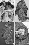
During the dissection, we found possible sign of pleural adhesion in her right-sided thorax, which is well correspondent to the findings on CT images (Fig. 3). The calcified PNs identified on the CT images were counter-checked by our dissection findings. Briefly, the calcified PNs were located in the apical segment of upper lobe, superior and posterior basal segments of lower lobe in the right lung. Next, we cut the nodules (#4 and #5) to examine their nature. The deposition of calcium seen in the center of nodules re-confirmed the radiopacities on CT images as dystrophic calcification (Fig. 3). Our results clearly show multiple calcified parenchymal nodules in the hilar region of the mummy's right lung. The radiological and anatomical information of calcified PNs is summarized in Table 1.
DISCUSSION
Most calcified PN on radiological images are clinically benign though some malignant diseases (e.g. primary or metastatic lung cancer), even if the chances are rare, also cause it. Based on the radiological pattern of calcified PN, we think that the current case might have been old healed granulomatous disease (calcified Ghon complex). Though intra-thoracic, diffuse nodular calcifications include granulomatous tissue response caused by various pathogens (Histoplasma capsulatum etc.) (8), pulmonary TB might be the first candidate in this case because it is one of the most common etiologies for calcified PN detected in the patients (9).
However, we should note that many other diseases such as infections, pulmonary metastases, pulmonary hamartoma, chronic hemorrhage, pneumoconiosis, carcinoid, amyloids, and other idiopathic disorders etc. also make calcified PN on radiological images (10). Differential diagnosis between them is therefore very important for the confirmation of our radiological diagnosis of the current case as pulmonary TB.
First of all, we do not regard this case as primary lung cancer because calcification in lung cancer is very rare; and if any, they show punctuate, amorphous and reticular patterns. We also rule out lung carcinoid because it is generally a central pulmonary tumor associated with dense amorphous calcification. If the current PNs were neoplasms, pulmonary metastases might be a better candidate for this case. However, as we could not get any radiological evidences for calcifications caused by primary cancer in the organs other than lung, calcified PNs in this study are not likely to be caused by pulmonary metastases.
As for pulmonary hamartoma (PH), we note that it appears as sharply outlined, solitary PN. PH generally associates popcorn-shaped round/oval PN with a central nidus and a laminated calcification pattern. The focal collection of fat that alternates with foci of calcification in PN is also a specific characteristic of PH on CT (9). Considering that the current case did not show such radiological patterns, we can rule out it easily from the diagnosis of current case. Amyloids also showed different pattern from the current case. As amyloids generally show well-defined 2-4 mm nodules with abnormal reticular opacities, diffusely dispersing in lung parenchyme (11), it can be excluded from the current diagnosis.
It is well known that calcified PN is also made by infectious diseases (e.g. healed disseminated histoplasmosis or miliary tuberculosis), hemosiderosis, pneumoconiosis, talcosis etc. However, we can rule out them without difficulties because they are very rare diseases, exhibiting the radiological patterns of multiple high-density, diffuse small calcification nodules throughout the lung parenchyma (910). Taken together, the general pattern observed in the current Korean mummy case was well matched with previous clinical findings reported from modern patients (1213). As far as our CT radiography and dissection results are concerned, the current case is likely to be the healed case of pulmonary tuberculosis (calcified Ghon complex), the first case identified among archaeologically obtained, pre-modern Korean (Joseon) samples.
In fact, revealing the earlier history of TB infection was a serious quest in paleopathology. Bartels (14) reported the evidence of spinal tuberculosis in a Neolithic skeleton discovered near Heidelberg, Germany in 1907. Spinal destructions possibly caused by Pott's disease were also identified in the ancient human bones from two different Italy sites, dated between 3500 and 4000 BC (1516). More recent studies on ancient tuberculosis cases were successfully summarized by Roberts and Manchester (17) in 2007 and Ortner (18) in 2003.
Although many pathological studies on archaeologically obtained human or animal samples worldwide have already been focused upon the evidences of ancient tuberculosis remained in them (19), similar researches were very rarely performed on the counterpart samples discovered in Korea. Only an exception was the ancient skeletal samples discovered in Nukdo archaeological site (Gyeongsangnam-do), which was dated to the first century BC. Upon the ancient Korean skeletons, Suzuki et al. (20) reported presumptive osteological sign of spinal tuberculosis having occurred in ancient Korean people. In fact, in the case, TV12 was collapsed wedge-shaped, and being fused with TV11.
Though this might be the first-ever report discussing about the presumptive tuberculosis case identified among ancient Korean remains, we should admit the technical limitation of this because the conclusion of Suzuki et al. (20) could have been derived from the simple observation on the skeletons. In fact, clear evidences on the TB infection affecting the lung parenchyme of ancient peoples therefore were not available yet in this country. In this regard, the current case is very significant to concerned researchers in Korea. As calcified PN in our report can be identified as an evident sign of pulmonary TB in a Joseon mummy, we show that the disease could have been also prevalent in pre-modern Korean society. Considering recent studies on ancient mycobacteria DNA worldwide have provided valuable addition to the knowledge of molecular evolution of the infectious disease, TB infecting human samples should be accumulated more in the future even from archaeological samples of Korea as well.


 XML Download