

 PDF
PDF ePub
ePub Citation
 Print
Print


INTRODUCTION
Although the JAK2 V617F gain-of-function mutation has a pivotal role in the diagnosis of Philadelphia chromosome-negative myeloproliferative neoplasms (Ph-MPN), it only occurs in 50%-60% in patients with essential thrombocythemia (ET) and primary myelofibrosis (PMF). Gain-of-function mutation in MPL, which is another molecular marker for ET and PMF, has been reported in 5%-10% of patients with these diseases with nonmutated JAK2 (1). Therefore, the diagnosis of other patients with findings suggestive of ET or PMF but negative for JAK2 and MPL mutations, must be based on the clinical exclusion of reactive thrombocytosis and histopathological examinations that focus on morphologic changes in megakaryocytes or bone marrow fibrosis. Histopathological evaluation is inherently susceptible to interobserver variation, requires an experienced hematopathologist, and lacks standardization. Therefore, it is very interesting that 60% to 88% of ET and PMF patients negative for JAK2 V617F and MPL mutations have been found to harbor novel mutations of the calreticulin gene, CALR (1, 2, 3, 4, 5, 6, 7, 8).
Calreticulin is a functionally complex Ca2+-binding protein that is localized primarily to the endoplasmic reticulum (9, 10, 11). All CALR mutations reported so far are located in exon 9 and are somatic insertions or deletions. There are two major variants: type 1 (L367fs*46), resulting from a 52-bp deletion, and type 2 (K385fs*47), from a 5-bp (TTGTC) insertion (1, 2, 3, 4, 5, 6, 7, 8). In particular, the clinical course of PMF patients with CALR alterations has been found to be more indolent than the courses of patients with the JAK2 V617F mutation and patients who are triple-negative (5, 6).
The aim of this study was to determine the prevalence, biological characteristics, and clinical correlations of these novel CALR mutations, in addition to the well-established JAK2 V617F and MPL mutations, in a single-center cohort of patients with ET and PMF.
MATERIALS AND METHODS
Patients
The study included 150 patients seen at the Asan Medical Center, Seoul, Korea from January 2003 to April 2013. There were 84 patients with ET, 50 patients with PMF, 7 patients with post-ET or post-polycythemia vera (PV) myelofibrosis, and 9 patients with other myeloid neoplasms with thrombocytosis, as follows: 4 refractory anemia with ring sideroblasts associated with marked thrombocytois, 1 refractory anemia with multilineage dysplasia, 1 myelodysplastic syndrome with isolated del(5q), 1 chronic myeloid leukemia, 1 myelodysplastic/myeloproliferative neoplasm, unclassifiable, and 1 acute myeloid leukemia. It should be stressed that the study patients, who were selected, were primarily those with available archived samples. Furthermore, patients with available data for JAK2 and MPL mutations were preferentially analyzed.
The median age of study patients was 60 yr (range, 19-90 yr); and 74 (49.3%) patients were male. ET, PMF, and other myeloid malignances were diagnosed based on 2008 World Health Organization (WHO) criteria. Post-ET or -PV myelofibrosis and leukemic transformation were also diagnosed according to 2008 WHO criteria. Major thromboembolic events at presentation or in the 2 preceding years or anytime during follow-up were recorded if definitely documented. Bone marrow aspirations and biopsies were independently reviewed by 2 hematopathologists who were blinded to the patient's mutation profile. Discrepant cases were resolved by consensus between the 2 hematopathologists. The clinical and laboratory parameters and cytogenetic findings at the time of initial presentation were reviewed. Karyotypes were designated as unfavorable based on the Dynamic International Prognostic Scoring System (DIPSS) risk categorization, as described previously; and included a complex karyotype (the presence of 3 or more distinct numeric or structural cytogenetic abnormalities) or 1 or 2 abnormalities that included +8, -7/7q-, i(17q), inv(3), -5/5q-, 12p- or 11q23 rearrangement (12).
Mutation and cytogenetic analysis
Study samples were either stored DNA extracted from peripheral blood or bone marrow of patients at initial presentation, or genomic DNA extracted from archived bone marrow smears according to a standard protocol and our laboratory's internal guidelines. The JAK2 V617F mutation was assessed using a polymerase chain reaction (PCR)-based amplification refractory mutation system, as previously described (13). The MPL W515L/K mutations were assessed by real-time PCR (Real-Q MPL W515L/K Screening Kit, BioSewoom Inc., Seoul, Korea) according to the manufacturer's instructions. All positive samples by real-time PCR were subsequently analyzed by Sanger sequencing. Exon 10 of MPL was amplified using the following primers: F, 5'-TTCTGTACATGAGCATTTCATCA-3'and R, 5'-GACAGGCTGTGTGTGTGTACCTCT-3'. The CALR mutations were assessed by PCR followed by Sanger sequencing. Exon 9 of CALR was amplified using the following primers: F, 5'-GAGGAGTTTGGCAACGAGAC-3'and R, 5'-AACCAAAATCCACCCCAAAT-3'. The PCR conditions consisted of initial denaturation step at 94℃ for 5 min; followed by 35 cycles of 94℃ for 45 sec, 57℃ for 30 sec, and 72℃ for 1 min; and a final extension step at 72℃ for 10 min. Purified PCR fragments were sequenced. Cytogenetic analysis was performed using conventional G-banding techniques.
Statistical analysis
Differences in the distributions of continuous variables between categories were analyzed using either the Wilcoxon rank-sum test or the Kruskal-Wallis test. Patient groups with nominal values were compared using the chi-square test or Fisher exact test. Overall survival (OS) was calculated from the date of diagnosis of ET or PMF to date of death (uncensored) or last contact (censored). Date of leukemic transformation replaced date of death (uncensored) to evaluate leukemia-free survival (LFS). OS and LFS were plotted using Kaplan-Meier curves and compared by a log-rank test. The Cox proportional hazard regression model was used for multivariate analysis. P values<0.05 were considered to indicate statistically significant differences. Statistical analyses were performed using MedCalc program version 12.4.0.0 (MedCalc Software, Acacialaan, Belgium).
RESULTS
Frequencies and distribution of mutations
CALR mutations were detected in 36 patients (25.5%) of patients with ET or PMF or post-ET or PV myelofibrosis. Among these patients, type 1 (L367fs*46), type 2 (K385fs*47) and other types of mutations were detected in 22 (61.1%), 10 (27.8%), and 4 (11.1%) patients, respectively. The type 1 mutation was found more frequently in patients with PMF than the type 2 mutation, with marginal statistical significance (P=0.076, Table 1).
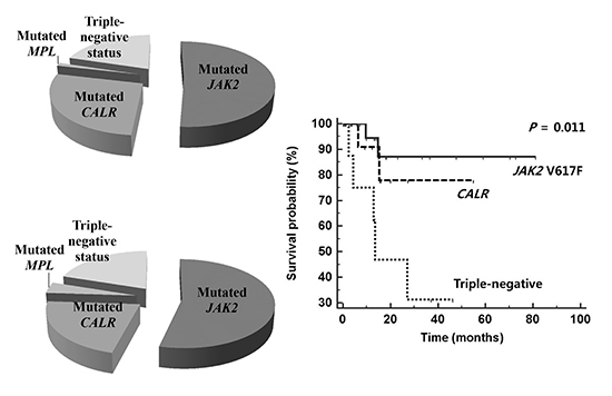
Of the 84 patients with ET, 43 (51.2%) harbored the JAK2 V617F mutation, 23 (27.4%) had CALR mutations, and 1 (1.2%) had MPL W515 mutation. Seventeen (20.2%) patients were negative for all 3 mutations (Fig. 1A). ET patients with CALR mutations accounted for 57.5% of patients that had nonmutated JAK2 and MPL. Of the ET patients with CALR mutations, 11 (47.8%) had type 1 mutations, 9 (39.1%) had type 2 mutations, and 3 (13.0%) had other types of mutations (Table 1).
Of the 50 patients with PMF, 27 (54.0%) harbored the JAK2 V617F mutation, 11 (22.0%) had CALR mutations, and 2 (4.0%) harbored the MPL W515 mutations. Ten (20.0%) patients were negative for all 3 mutations (Fig. 1B). PMF patients with CALR mutations accounted for 52.4% of the patients with nonmutated JAK2 and MPL. Of the patients with CALR mutations, 9 (81.8%) patients had type 1 mutations, 1 (9.1%) patient had a type 2 mutation, and 1 (9.1%) patient had another type of mutation (Table 1).
Of the 7 patients with post-ET or PV myelofibrosis, 5 (71.4%) harbored the JAK2 V617F mutation and the remaining 2 (28.6%) had CALR mutations. The 2 CALR mutations were only found in patients with post-ET myelofibrosis (Table 1), whereas the 2 patients with post-PV myelofibrosis who were included in this study had the JAK2 V617F mutation. These 3 mutations (CALR/JAK2/MPL) were mutually exclusive and were never detected in patients with other myeloid neoplasms with thrombocytosis.
Clinical, hematologic and molecular correlates
Tables 2 and 3 summarize the presenting clinical characteristics of study patients stratified by mutational status as follows: mutated CALR, JAK2, MPL and triple-negative. Univariate analysis of patients with ET found that CALR mutations were associated with a higher platelet count (P=0.006), lower leukocyte count (P=0.035) and higher frequency of cytogenetic abnormalities (P=0.017), compared to the JAK2 V617F mutation. CALR mutations were associated with a higher lactate dehydrogenase (LD) level (P=0.004) and lower leukocyte count (P=0.039) compared with triple-negative status. A triple-negative status was associated with a lower hemoglobin level (P=0.021), lower LD level (P=0.025) and higher frequency of cytogenetic abnormalities (P=0.049), compared with the JAK2 mutation (Table 2). Thromboembolic events seemed to be more frequent in patients with the JAK2 V617F mutation compared to patients with CALR mutation or triple-negative status, although statistical significance was not achieved. Neither post-ET myelofibrosis or nor leukemic transformation was documented during follow-up.
Of patients with PMF, CALR mutations were associated with a higher platelet count (P=0.035), and there was a trend toward lower frequency of cytogenetic abnormalities (P=0.053), compared to the JAK2 V617F mutation. CALR mutations were also associated with a higher platelet count (P=0.020) compared with triple-negative status. A triple-negative status was associated with a lower hemoglobin level (P=0.001) and lower LD level (P=0.037), compared to the JAK2 V617F mutation. CALR mutations had lower frequency of leukemic transformation compared to the JAK2 V617F mutation or triple-negative status, although statistical significance was not achieved (Table 3).
Prognostic impacts of mutations
The median follow-ups of the study population were 29.8 months (range, 0.2-120.2 months) for ET patients and 13.7 months (range, 0.2-81.2 months) for PMF patients. Four (4.8%) ET patients died, and 9 (18.0%) PMF patients died. The mortality rates of PMF patients with the JAK2 V617F mutation, CALR mutations, and triple-negative status were 7.4% (2/27), 18.2% (2/11), and 50.0% (5/10), respectively.
OS and LFS times were analyzed according to the various parameters, including mutational status. Mutational status, including CALR and JAK2 V617F mutations, had no significant prognostic impact on patients with ET (P=0.124). However, among PMF patients, the median OS time of those with CALR mutations, the JAK2 V617F mutation, and triple-negative status was 15.7, 14.3, and 13.0 months, respectively. Univariate analysis found significant survival differences between patients with JAK2 and CALR mutations and triple-negative patients (P=0.011 for OS and P=0.026 for LFS). Triple-negative status was associated with shorter OS than JAK2 V617F mutations (hazards ratio [HR], 7.7; 95% confidence interval [CI], 1.4-42.9) and CALR mutations (HR, 3.6; 95% CI, 0.8-16.0; Fig. 2A). Triple-negative status was also associated with shorter LFS than JAK2 V617F mutations (HR, 4.3; 95% CI, 1.0-17.7) and CALR mutations (HR, 2.9; 95% CI, 0.8-10.8; Fig. 2B). Among the other clinical parameters, univariate analysis found that an intermediate-2 or high DIPSS score was associated with significantly shorter OS (HR, 10.8; 95% CI, 2.4-49.5; P=0.0001) and LFS (HR, 6.5; 95% CI, 1.8-23.1; P=0.0001) than an intermediate-1 or low score. An unfavorable karyotype was associated with shorter LFS (HR, 4.0; 95% CI, 1.1-14.7; P=0.005), but not shorter OS.
By multivariate analysis, triple-negative status (HR, 7.0; 95% CI, 1.6-31.1, P=0.01) and intermediate-2 or high DIPSS score (HR, 22.4; 95% CI, 2.6-196.0, P=0.005) were associated with shorter OS times. Triple-negative status (HR, 6.3; 95% CI, 1.8-22.0, P=0.004), intermediate-2 or high DIPSS score (HR, 9.7; 95% CI, 2.5-37.3, P=0.001), and unfavorable karyotypes (HR, 4.5; 95% CI, 1.4-14.6, P=0.013) were associated with shorter LFS.
DISCUSSION
The most consistent finding of this study was that among ET and PMF patients, those with CALR mutations had significantly higher platelet counts than those with the JAK2 V617F mutation. Previous studies revealed that CALR mutations were not found in patients with PV (1, 2), and that CALR mutations were found in few, if any, patients with myeloid neoplasm with thombocytosis (1). Both these observations and our results are consistent with a recent study that used measurements of the levels of calreticulin RNA and immunostaining by an antibody specific for mutated CALR. It found that calreticulin expression is restricted to megakaryocytes (14). In the endoplasmic reticulum (ER), calreticulin has important functions in directing the proper conformation of proteins and glycoproteins, as well as in homeostatic control of cytosolic and ER calcium levels (9, 10, 11). Because transient ER stress activation is thought to trigger the apoptotic-like phase of the thrombopoiesis process (11), the effects of calreticulin as a calcium-buffering chaperone of the ER and ultimately as a contributor to platelet production might vary, depending on the presence or absence of a CALR mutation. The key region during the molecular pathogenesis of Ph-MPN is the acidic C-domain that terminates in a KDEL sequence, and which is involved in the homeostasis of cellular calcium and is lost in mutated CALR. The absence of this sequence in mutated CALR results in altered intracellular localization and impairment of the calcium-binding function (1, 2, 15).
Overall, the other findings of our study also confirm the results of previous studies. Our study found that the frequency of CALR mutations was 27.4% in patients with ET and 22.0% in patients with PMF. The reported frequencies of CALR mutations range from 15.5% to 33% in ET patients and from 22.7% to 25% in PMF patients (3, 4, 5, 6, 7, 8). The lower leukocyte counts in the CALR-mutated ET patients in this study, compared to the JAK2-mutated ET patients, has also been reported in previous studies (3, 4, 7, 8). Among patients with PMF, the triple-negative patients in our study had lower hemoglobin levels and trended toward higher prevalence of cytogenetic abnormalities and acute leukemic transformation than the JAK2- and CALR-mutated PMF patients. Ultimately, the triple-negative PMF patients had the shortest OS and LFS. These findings for triple-negative patients have also been reported by previous studies, and corroborate the adverse effects that triple-negative mutations had on the survival times of patients with PMF in our study (5, 6, 8). It is not surprising, therefore, that Tefferi et al. have emphasized that triple-negative PMF should be thought of a molecularly high-risk disease (5).
Molecular variations resulting from genetic alterations may be highly diverse between different ethnicities. Since the studies referred to in our discussion were principally performed in Western countries, a review of studies on Asian populations is needed. We found 4 very recent reports in the PubMed database on CALR mutations in studies of Chinese populations (16, 17, 18, 19). On the whole, the results of these Chinese studies are in line with the results of the Western studies, regarding the frequencies of mutations and associated clinical characteristics and prognostic impacts. Of note, the results of a single study on Chinese PMF patients reported by Li et al. indicate that CALR mutations may have different effects in different populations (19). First, their study found that 43.8% of patients with no detectable mutations in JAK2 or MPL had CALR mutations. This frequency is substantially lower than the frequencies for PMF patients of predominantly European descent, which range from 69.4% to 74.2% (5, 6, 8), but is similar to the frequency in our study (52.4% of JAK2- or MPL-unmutated PMF patients). The reasons accounting for the discrepant findings include the different characteristics of the study patients, different sensitivities of the methods used, and ethnicity-based differences in genetic profiles. The last reason may be a reasonable explanation, because of the slightly lower frequencies of JAK2 V617F and MPL mutations in Asian patients compared to Western patients. The frequencies of the JAK2 V617F mutation in a Chinese study (19) and our study were 50% and 54%, respectively, versus 58% and 64.7% in Western studies (5, 6). The frequency of MPL mutations in a Chinese study (19) and our study was 3% and 4%, respectively, versus 8.3% and 7% in Western studies (5, 8). Second, the ratio between type 1 and type 2 CALR mutations was reversed in the Chinese PMF patients (32% were type 1 and 64% were type 2 mutations) compared with PMF patients of predominantly European descent (19). A large study of Italian PMF patients found frequencies of type 1 and type 2 mutations of 72% and 16%, respectively (6). Our results also found a predominance of type 1 mutation in PMF patients compared with ET patients. Therefore, the predominance of the CALR type 2 mutation in Chinese PMF patients should be confirmed in a larger Asian cohort.
The identification of disease-specific mutations justifies, in certain cases, the use of mutation analysis for determining the diagnosis, prognosis and assessment of response to treatment. CALR mutations are frequent in JAK2/MPL-unmutated ET/PMF and therefore are important clonal markers for such cases. Therefore, Tefferi et al. have urged that these mutations should be included as clonal markers, along with JAK2 and MPL mutations, in the list of major diagnostic criteria for ET and PMF in the upcoming WHO classification system (20).
The major limitations of this study include the relatively low numbers of patients in our cohort and relatively short follow-up period. This might be major causes for the lack of data demonstrating the clinical relevance of CALR mutations, such as longer thromboembolic event-free survival and lower rate of thrombotic events for ET patients with CALR mutations than for patients with JAK2 V617F mutations (3, 4, 8), or better OS for PMF patients with CALR mutations compared to PMF patients with JAK2 V617F mutations (5, 6, 8). In addition, type 1 and type 2 CALR mutations have manifested as distinctly different clinical phenotypes. The type 1 mutation was found to cause in vitro cytokine-independent growth as a result of its activation of the signal transducer and activator of transcription 5, while the type 2 mutation was found to be associated with increased platelet counts and probably has an important role in the maturation of megakaryocytes (1, 7). Furthermore, in the previously mentioned study on Chinese PMF patients, the unfavorable prognostic impact of CALR mutations was limited to patients with type 2 mutations, and type 2 mutations or no detectable mutation was an independent unfavorable prognostic factor by multivariate analysis (19). In our study, we could not detect differences between the 2 types of CALR mutations, mainly because of the relatively small number of study patients.
Although a larger study cohort is definitely needed to validate the results of this study, our results should not be underestimated because, to the best of our knowledge, this is the first study that has investigated the frequency of CALR/JAK2/MPL mutations and related biologic and clinical features in Korean Ph-MPN patients. Recently, two Korean studies on CALR mutations were reported. However, the clinical usefulness of these studies seems to be limited, either by lack of prognostic data (21) or exclusion of MPL mutation and small size of each subgroup (22). Moreover, CALR mutations will probably be included in the major criteria for ET and PMF of the new WHO classification system (20). We have confirmed that CALR mutations occur in Korean patients with Ph-MPN that lack JAK2 V617F or MPL mutations. Therefore, diagnosis of ET or PMF can now be facilitated by analyzing CALR mutations in Korean patients. In addition to the diagnostic value of mutation profiling, it is also useful for detecting high-risk PMF patients who are triple-negative for mutations.
In conclusion, we demonstrated that CALR mutations characterize a subset of Korean Ph-MPN patients who have distinct phenotypes similar to those reported for patients in Western countries. Because molecular markers are still unknown for approximately 50% of JAK2- and MPL-unmutated cases, morphological examination of bone marrow remains the cornerstone for accurate diagnosis for a substantial number of Korean patients suspected of having Ph-MPN. CALR mutations provide additional information on clinical outcomes, especially for patients with PMF.


 XML Download