

 PDF
PDF ePub
ePub Citation
 Print
Print


INTRODUCTION
Traditionally, colorectal carcinoma (CRC) has been graded as well-differentiated (WD), moderately differentiated (MD), poorly differentiated (PD), and undifferentiated on the basis of the percentage of gland formation (1). Although histologic grading of tumor differentiation has been shown repeatedly by multivariate analysis to be a stage-independent prognostic factor, in fact, this type of grading poses practical problems for pathologists and has limitations as a useful tool for clinicians. For pathologists, a significant degree of interobserver variability exists (2, 3). Furthermore, applying the grading to carcinomas of unusual types of tumor, such as mucinous, signet ring cell, medullary, and micropapillary carcinomas is of questionable practical utility and remains debatable. For clinicians, the difference in outcome between patients with well-differentiated and moderately differentiated tumors is not as clear as the difference between well or moderately differentiated tumors and poorly differentiated tumors (4). Moreover, because there is only a small fraction of poorly differentiated tumors by conventional histologic grade, inevitably only a small fraction of patients is provided reliable prognostic information.
A recently proposed histologic approach to the assessment of tumor aggressiveness considers tumor budding. European Society Medical Oncology (ESMO) guidelines (2012) recognized tumor budding as a "potential prognostic factor" for curatively resected CRC, but the standardization of tumor budding assessment remains a work-in-progress (5, 6). A more recent study by Ueno et al. (7) focused on the histologic features of poorly differentiated cluster (PDC), defined as a solid cancer cell nest comprising ≥ 5 cancer cells and lacking a gland-like structure. Ueno et al. (7) maintained that the novel grading system based on number of PDCs is an accurate, reproducible and predictable grading that is easier and more objective than conventional histologic grading or tumor budding grading. Although this new grading system has been shown to have simplicity, as well as advantages of reproducibility and a robust prognostic value in some reports, there remains a need for continued validation of the reliability of the novel grading system (8, 9, 10, 11).
It is well known that microsatellite instability (MSI) is associated with specific and variable histological features. Tumors with MSI-high (MSI-H) have a pushing margin, an exophytic/polypoid growth pattern, lymphocytosis, and signet ring cells. Furthermore, tumors with MSI-H are more frequently heterogeneous, mucinous, or medullary type or poorly differentiated (12). Because PDCs-based grade is a quantitative method, it has the advantage of being capable of application to CRCs with these variable morphologies, unlike conventional histologic grade, in which it is difficult to integrate diverse morphologic patterns.
In this study, we aimed to investigate the association between variable clinicopathological parameters and PDCs-based grade and to analyze the prognostic significance of the novel grading system. In addition, we examined the relationship between the novel grading system and MSI.
MATERIALS AND METHODS
Patients and histopathologic evaluation
A total of 235 patients who underwent a standard radical colorectal surgery and regional lymphadenectomy for primary colorectal adenocarcinoma at Kangnam Sacred Heart Hospital in Seoul between September 2008 and December 2012 were retrospectively analyzed. Cases with preoperative neoadjuvant chemoradiotherapy, multiple primary cancers such as synchronous and metachronous tumors, and non-radical surgery such as local resection or polypectomy were excluded. Furthermore, patients who had invalid MSI results were also excluded. Ultimately, 201 patients were eligible, and demographic, clinical and follow-up data were collected from the patients' medical records. Proximal tumors referred to tumors located at the cecum, ascending colon, hepatic flexure and transverse colon.
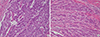
Hematoxylin-eosin-stained slides of all tumor samples were reviewed by two pathologists who were blinded to the patient's clinical information, and the tumors were staged according to the seventh edition of the American Joint Commission on Cancer (AJCC) cancer staging system (13). For each case, histologic grading was performed based on glandular differentiation according to the World Health Organization (WHO) criteria (1) and on counting PDCs according to the previously described criteria by Ueno et al. (7, 9). In brief, PDCs were defined as cancer cell clusters in the tumor stroma composed of ≥5 cancer cells and lacking a gland-like structure, regardless of the size of the cluster (Fig. 1). More specifically, with regard to the assessment of mucinous carcinoma, malignant clusters with no tubular formation infiltrating the stroma with minimal extracellular mucin were classified as PDCs. Cancer cell clusters within a large mucin pool (i.e. mucinous lake) were not classified as PDCs. Also, clusters with invasive micropapillary pattern or signet ring cells were accepted as PDCs if they had clustering and no tubular formation. To quantify these PDCs, the whole tumor including its advancing edge was scanned at lower-power magnification to identify the area with the highest number of PDCs. The clusters were counted using a ×20 objective lens (i.e. a microscopic field with a major axis of 1 mm). Tumors with <5, 5-9, and ≥10 clusters were classified as grade 1 (G1), grade 2 (G2) and grade 3 (G3), respectively. Other histological findings, including lymphovascular invasion (LVI), perineural invasion (PNI), mucin production and tumor budding were also assessed. Tumor budding was defined as isolated single cancer cells or as cancer clusters with <5 cancer cells observed in the desmoplastic stroma of the actively invasive region. After choosing one field in which budding was most intensive, a budding count was made using the ×20 objective lens (0.785 mm2). Budding was divided into two groups: low grade (counts of 0-9) and high grade (counts of 10 or more) as previously described (14, 15).
Molecular analysis
Genomic DNA was obtained from formalin-fixed paraffin-embedded non-neoplastic and neoplastic colorectal tissue and was purified using the QIAamp DNA Mini Kit (Qiagen, Hilden, Germany) according to the manufacturer's protocol. Analysis of MSI status was based on the multiplex amplification of the microsatellites (BAT25, BAT26, D2S123, D5S346, and D17S250) recommended by a National Cancer Institute (NCI) consensus group (16). POP-7 polymer solution (Applied Biosystems, Foster City, CA, USA) was used for electrophoresis on the ABI Prism® 3100 Genetic Analyzer (Applied Biosystems). Tumors were classified as MSI-high when at least two of the five loci showed MSI and as MSI-low (MSI-L) when only one locus showed MSI. If none of the five microsatellite sequences were mutated, the tumor was classified as microsatellite stable (MSS). Patients with MSS and MSI-L tumors were defined as the low MSI group in this analysis, and those with MSI-H tumors were defined as the high MSI group (17).
Statistical analysis
The association between PDCs-based grading and other clinicopathologic characteristics (age, sex, tumor site, tumor size, WHO differentiation, TNM stage, LVI, PNI, and tumor budding) and MSI status were analyzed by the chi-square test, Fisher's exact test, logistic regression analysis and Kruskal-Wallis test. Survival curves were calculated using the Kaplan-Meier method, and differences between curves were evaluated using the log-rank test. The Cox proportional hazards regression analysis was used to determine the impact of histologic parameters on disease-specific survival (DSS) and disease-free survival (DFS). All statistical analyses were performed using the Statistical Package for Social Sciences software program, version 21.0 (SPSS Inc., Chicago, IL, USA), and the results were considered statistically significant when the P value was ≤0.05.
RESULTS
Correlations between PDCs-based grade and clinicopathologic parameters
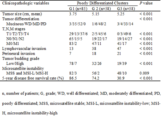
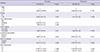
The mean age of study patients was 62.2 yr of which 108 (53.7%) were male. The median follow-up period was 34.5 months. One hundred twenty-six (62.7%) tumors were located in the colon and 75 (37.3%) in the rectum. Histologically, 7 mucinous adenocarcinomas were noted, and 194 non-mucinous adenocarcinomas were classified as WD (23.1%), MD (68.6%), or PD (8%) (Table 1).
Based on the number of PDCs, 85, 58, and 58 tumors were classified as G1 (42.3%), G2 (28.9%), and G3 (28.9%), respectively. PDCs-based grade was significantly associated with tumor size, conventional histologic grade, T stage, N stage, M stage, LVI, PNI, and frequent tumor budding (all P<0.001). With regard to the disease progression of pM0 tumors, PDCs-based grading was closely correlated with the incidence of local recurrence and distant metastasis (P<0.001).
Prognostic impact of PDCs-based grading system
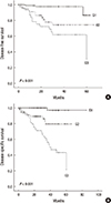
The 5-yr DFS rate was 86.5% for the G1 tumor group and extremely favorable, whereas it was 74.2% for the G2 tumor group and 30.9% for the G3 tumor group (P<0.001). The 5-yr DSS rates for those same groups were 96.7%, 79.4% and 21.6%, respectively (P<0.001) (Fig. 2).
Univariate analysis revealed that PDCs-based grade, T stage (≥T3), N stage (≥N1), LVI, PNI, and tumor budding grade were poor prognostic factors for DFS (P = 0.001, 0.009, 0.002, 0.003, 0.006, and 0.001, respectively) (Table 2). By multivariate analysis, PDCs-based grade was selected as an independent prognostic parameter (P=0.022; hazard ratio [HR]=3.709 [G2], 7.461 [G3]). In a similar manner (not shown), M stage (M1) and the novel grade had a significant impact on DSS by multivariate analysis (P<0.001 and 0.005, respectively; HR=6.119 [G2], 19.056 [G3]). Grade based on differentiation did not have a prognostic significance.
Relationship between microsatellite instability and PDCs-based grade
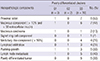
Of 201 total tumors, 15 were classified as MSI-H (7.5%), of which 8 were located proximally (53%). PDCs-based grade was associated with MSI-H (P= 0.009) (Table 1). In particular, G3 tumors were significantly correlated with MSI-H tumors compared to G1 and G2 tumors (P=0.002, odds radio [OR]=5.750).
We reviewed histologically the tumors with MSI-H and summarized each characteristic according to PDCs-based grade, as shown in Table 3. These tumors tended to produce mucin (60%; P=0.011). One of the two mucinous carcinomas showed signet ring cell components as PDCs and was classified as G3. Typical medullary carcinoma was not noted, but there were four tumors (27%) with a medullary-like component of >10%; all 4 were classified as G3 (Fig. 3). Peritumoral or intratumoral lymphoid infiltrates and a pushing growth pattern were found in 27% and 33% of tumors, respectively. Six tumors had lymph node metastasis (40%), 5 of which were classified as G3. Additionally, 5 out of 16 poorly differentiated tumors according to conventional histologic grading frequently showed MSI-H, and these were all classified as G3. Multivariate analysis demonstrated that only mucin production was independently associated with MSI-H (P<0.001, OR=11.437).
DISCUSSION
Histologic grading endorsed by the WHO and AJCC is one of the most widely used pathologic variables in CRC, but it is also one of the most difficult to define accurately. This difficulty is primarily a result of the heterogeneous degree of differentiation of CRC, which is a distinctive characteristic of the tumor. Occasionally, CRC shows less differentiation at the leading edge where the tumor is most aggressive than at the superficial component. However, no standard international criteria have been established for judging whether grading should be diagnosed on the basis of the predominant pattern of differentiation or on the area of least differentiation. Therefore, increasing subjectivity and interobserver disagreement are inherent in the grading system. Furthermore, the current grading system cannot be applied to all CRC types, such as medullary and mucinous carcinomas, and there has been a recommendation that these tumors be ungraded (18). For the last two decades, therefore, the issue of the current differentiation-based grading system being a less objective and suboptimal tool with inadequacy in predicting behavior has been raised.
Recently, Ueno et al. (7) suggested a novel grading system based on the number of PDCs and demonstrated the superiority of this grading system in terms of interobserver agreement and prognostic power. This method has subsequently been followed, and its reproducibility has been verified by Barresi et al. (8, 10, 11). However, the studies did not include mucinous carcinoma and excluded mucinous areas on examination of heterogeneous CRCs because of the insufficient description of the PDC counting method in the original paper. On the reference to the following report of Ueno et al. (9) with more specified explanation, we included mucinous carcinoma in this study and examined mucinous areas in CRCs with mucinous components. Also, grading in CRCs with heterogeneous morphology, such as micropapillary and medullary-like components could be coherently approached. Consistent with the results in previous reports, our results showed that PDCs-based grade was a robust prognostic factor and is significantly correlated with clinicopathologic parameters associated with disease progression, T stage, N stage, M stage, LVI, PNI, and tumor budding grade.
With regard to morphology and identification, although PDCs show a histologic similarity to tumor budding in terms of loss of gland formation, some histopathologies are distinguishable. According to the definition of tumor budding (composed of <5 cells), there is practical difficulty in precisely identifying and counting fairly small tumor budding or single cancer cell. Tumor budding is frequently observed in the actively invasive frontal region, whereas PDCs often appear within a tumor and at the advancing edge of the tumor. In addition, we observed that tumor budding almost always accompanies desmoplasia, whereas PDCs might show less desmoplastic reactions and could be counted in more diverse morphologic backgrounds, such as mucinous, signet ring cells and micropapillary- and medullary-like features. Therefore, analyzing PDCs was more appropriate than analyzing tumor budding for comprehensive application to the grading of tumors with heterogeneous morphology.
Interestingly, we found that the new grading system has a more proportionate distribution of tumors classified in each category. By conventional grading, 68.6% and 23.1% of cases in this study were MD and WD, respectively and only just 8% were PD. Using the novel grading system, 29% were G2 tumors, the percentage of G1 tumors increased to 42% and the percentage of G3 tumors increased to 29%. Moreover, each group showed distinct survival differences with statistical significance. Therefore, we believe that this grading system would give a greater number of patients more reliable prognostic information.
To our knowledge, the present study is the first to evaluate a relationship between PDCs-based grade and MSI status, which is a potential element for the molecular classification of CRC. It is well known that tumors with MSI-H show considerably inconsistent histologic characteristics, with mucinous or medullary features and pushing growth versus poorly differentiated or signet ring cells feature. These specific histological subtypes could make interpretation of a relationship between MSI and tumor grade both difficult and confusing. In this study, G3 tumors classified by the PDCs-based grading system correlated with MSI-H. Further, poorly differentiated tumors according to the WHO grade showed significantly frequent MSI-H (P=0.002). However, the budding grade showing some morphologic similarities with PDCs was not correlated with MSI-H (P=0.334). An association between MSI status and tumor budding has been previously described. Zlobec et al. (17) and Jass et al. (19) reported that tumor budding was inversely correlated with MSI-H. However, they considered ≥6 tumor buds/0.65 mm2 as a high tumor budding grade, compared to the ≥10 tumor buds/0.785 mm2 field of vision considered in our study. There is no current standard for the "optimal" threshold of high-grade tumor budding, but the relatively low cut-off point used by Zlobec et al. (17) and Jass et al. (19) should be considered. In addition, the morphological diversity of PDCs compared to tumor budding might be another factor causing the difference in correlation with MSI-H status. In our study, 4 tumors with medullary-like components were all classified as G3 and showed MSI-H, but they were low grade according to tumor budding grade (Fig. 3). Typical medullary carcinoma with large sheets would be classified as low grade according to the PDCs-based grade, but we observed tumors with many PDCs in a medullary-like background. Lastly, it should be considered that we examined mucinous areas in CRCs with mucinous components. In our study, mucin production was an independent predictive factor of MSI-H.
In this cohort, the frequency of the MSI-H genotype was 7.5%, which is lower than in studies of Caucasians but similar to other studies of Chinese and Koreans (17, 20, 21, 22). In addition, MSI-H cancers in this study showed a male predominance (2:1) in contrast to studies of Caucasians, in which there is a marked female predominance (about 3:1) (12). Because discrepancies based on ethnicity and/or environmental effects might affect the association of PDCs-based tumor grade and MSI status, subsequent studies in variable cohorts would be valuable.
Despite having several positive findings, our study had some limitations. First, this was a single-institution-based retrospective study. Second, although the study was based on a large population size, the incidence of specific histologic subtypes such as pure mucinous carcinoma and medullary carcinoma was very low. Third, the lack of information regarding postoperative chemotherapy/radiation treatment and the short follow-up period may have obscured the prognostic analysis.
In conclusion, the present study reveals that the novel CRC grading system based on PDC counting would provide a greater number of patient valuable prognostic information and is feasible for CRCs with heterogeneous morphology. This grading system deserves continued verification for acceptance as the optimal and substitutable method of CRC tumor grading. The association between G3 tumors and MSI-H suggests that MSI may play a role in forming PDCs. However, we propose that this result should be further tested in a larger cohort with specific histologic subtypes in order more clearly to understand the nature of PDCs.

 XML Download