

 PDF
PDF ePub
ePub Citation
 Print
Print


INTRODUCTION
Radiation-induced enteritis is a common and serious complication after abdominal or pelvic irradiation for gastrointestinal, gynecologic and urologic cancer (1, 2, 3). More than 70% of patients receiving pelvic radiation therapy suffered from bowel symptoms caused by inflammatory changes, and 50% of them developed into chronic bowel disease with permanent intestinal changes (4). Therefore, the methods to prevent or decrease the occurrence of the acute radiation-induced enteritis may be the one of the important issues to reduce the chronic toxicity. Although the more conformal irradiation techniques such as three-dimensional radiation therapy or intensity-modulated radiation therapy allowed a significant improvement in normal organ sparing, the radiation-induced enteropathy still has been reported with high incidence rate in patients receiving pelvic irradiation (5). While the highly accurate irradiation techniques can reduce the dose to the bowels, they may increase the intestinal volume exposed to relatively lower radiation dose because of multiple directions of radiation beams (6). The advanced irradiation techniques may be an insufficient solution of radiation-induced intestinal injury.
Probiotics are defined as live microorganisms such as Lactobacillus acidophilus that exert specific favorable effects on the host. Continuous intake of them is thought to restore bowel microflora to optimal levels and reinforce the intestinal barrier capacity. Probiotics have been reported to prevent radiation-induced enteropathy and diarrhea (7). Experimental animal studies of acute radiation-induced enteritis have revealed that probiotics have a positive role on the mucosal integrity (8, 9). However, the effect of probiotics for radiation-induced diarrhea is still controversial issue in clinical studies (10, 11, 12).
The morphological damages of acute radiation-induced enteritis were known as architectural changes of intestinal mucosa such as villus shortening by apoptosis (13, 14). In the present study, morphometric analysis of bowels was performed in rats to evaluate the effect of L. acidophilus on the morphological changes of intestinal mucosa by irradiation and their correlation with radiation dose.
MATERIALS AND METHODS
Animals and probiotics
A total of 48 adult male Sprague-Dawley rats, each weighing 300-350 g, were enrolled in this study. The rats were housed in a standard wire cages with a constant temperature of 22±2℃, with 12 hr lighting. They were fed with standard chow diet and UV-sterilized water.
All rats were randomly assigned to two groups; L. acidophilus group and placebo group. L. acidophilus group received 2 mL of solution that contained 1.0×10⁸ colony-forming units (CFU) of L. acidophilus daily by feeding canula for 10 days. The probiotic solution was achieved by dissolving freeze-dried L. acidophilus powder in water. Placebo group was given only 2 mL of water using same methods during same period as L. acidophilus group.
Irradiation
Each group was randomly allocated to three subgroups of 8 rats in each subgroup on the seventh day after feeding L. acidophilus or placebo solution. All animals were anesthetized with an intraperitoneal injection of 100 mg/kg of ketamine hydrochloride. Then four rats at a time were restrained and taped by the tail on 1 cm-thicked acrylic plate with supine position and covered by another acrylic plate with same thickness. Each subgroup was irradiated with single dose of 10, 15, and 20 Gy, respectively, using 6 MV photon beam. The radiation was administered to abdomino-pelvic area at 3 cm depth through anterior-posterior/posterior-anterior fields using linear accelerator (Clinac 21EX, Varian Medical System, Inc., Palo Alto, CA, USA). Dose rate of the irradiation was 1.04 Gy per minute.
Histologic evaluation
Each group was given L. acidophilus or placebo solution until third day after irradiation. All rats were sacrificed by carbon dioxide asphyxiation 6 hr after the last feeding the solutions and underwent laparotomy immediately. One-centimeter segments of jejunum, ileum and colon were resected and washed with saline solution. Tissue samples were fixed in 10% buffered formalin for 24 hr and embedded in paraffin. The sections were cut at 5 µm and stained with hematoxylin and eosin. The mucosal thickness (from the base of muscularis mucosa to the tip of the villus) and villus height (from the base to the tip of the villus) of each segment were measured at four separate microscopic fields using light microscopy at a 100×magnification and recorded as the mean values of them. All morphometric measurements were performed in a blind fashion without the knowledge of the solution groups.
Statistical analysis
The changes of mucosal thickness and villus height according to the radiation dose were evaluated by Kruskal-Wallis test. The comparisons of the parameters between L. acidophilus group and placebo group were analyzed by Mann-Whitney U-test. A statistical analysis was performed using SPSS, version 19.0 (IBM, Somers, NY, USA).
RESULTS
Mortality
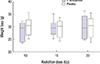
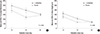
There were total 3 deaths from diarrhea in the subgroups irradiated with 20 Gy; one rat in L. acidophilus group and two in placebo group. No mortality was observed in the rats irradiated with 10 Gy or 15 Gy in both groups. Statistical comparison to evaluate mortality was not performed because the number of the rats was too small. Mean weight loss of 29 g (range, 23 to 37 g) was observed in the whole rats and, there was no statistically significant difference in weight loss between two groups (P=0.942). The comparison of weight loss by radiation dose between the solution groups is displayed in Fig. 1.
Jejunum
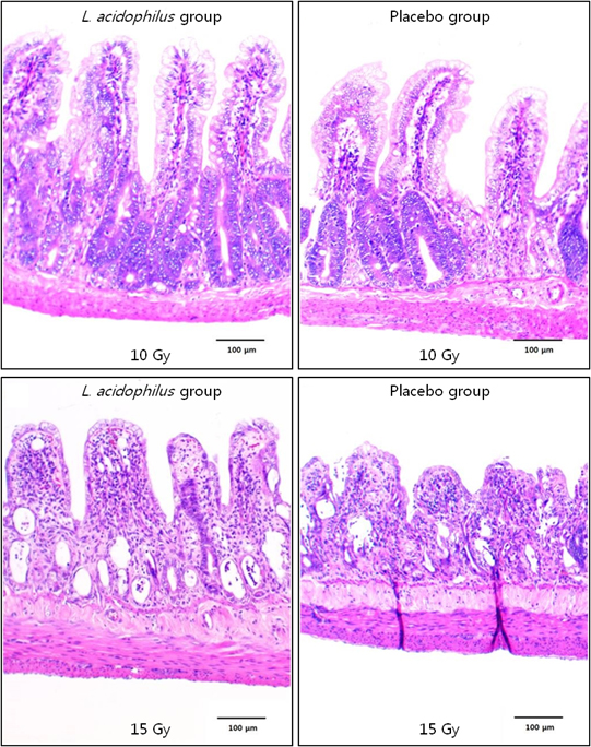
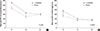
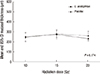
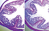
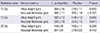
Mucosal thickness and villous height of jejunum decreased significantly as the radiation dose increased (P<0.001 for both; Fig. 2). In the comparison with placebo group, mucosal thickness and villous height of jejunum were significantly taller in L. acidophilus group after 10 Gy of irradiation (P<0.001 and P=0.003, respectively; Table 1). Histologic findings of jejunum after 10 Gy irradiation are displayed in Fig. 3A and B. In the rats were irradiated with 15 Gy, thickness of jejunal mucosa in L. acidophilus group were significantly greater than that in placebo group (P=0.015). However, the analysis of villus height of jejunum showed a marginally significant difference between two groups (P=0.065). There was no significant difference of the two morphometric variables between the solution groups in jejunum of the rats received 20 Gy of irradiation.
Ileum
With the higher dose of radiation compared with the lower dose, there were significant decreases in mucosal thickness and villous height of ileum (P<0.001 for both; Fig. 4). There were significantly greater thickness of mucosa and height of villi in L. acidophilus group irradiated with 10 Gy than those in placebo group (P=0.028 and P=0.010, respectively; Table 2). The two histologic parameters of ileal segments after 15 Gy irradiation also presented significantly higher values in L. acidophilus group (P<0.001 for both). Fig. 3C and D show the histologic features in ileum after 15 Gy of irradiation. The rats irradiated with 20 Gy had no significant difference in morphometric analysis of ileal mucosa between L. acidophilus group and placebo group.
Colon
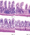
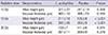
There was no significant change in mucosal thickness of colon by increasing radiation dose (P=0.174; Fig. 5). Table 3 shows that the thickness of colon mucosa did not differ between L. acidophilus group and placebo group after irradiation with the dose of 10, 15, and 20 Gy. Histologic findings in large bowel of rats is shown in Fig. 6.
DISCUSSION
The results of this trial show at least marginally significant benefit of L. acidophilus for decreasing the mucosal damages of small bowels induced by radiation of 10 or 15 Gy in rats. There was no difference in mucosal morphometrics of small intestines between L. acidophilus group and placebo group after 20 Gy of irradiation. Cell necrosis and mucosal ulceration of small intestine were reported to be evoked mainly by radiation of more than 20 Gy (15). Therefore, high dose irradiation more than 20 Gy might devastate the mucosa of small bowel and interfere with them to be restored by L. acidophilus.
There were many reports with a wide variation in the dose of probiotics and they showed that administering probiotics at a high dose of more than 1.0×10¹⁰ CFU per day is safe in rats (16). The minimum effective dose of probiotics is usually considered as 1.0×10⁸-109 CFU per day (17). Probiotics were also reported to be present at high concentration on the seventh day of the daily ingestion because they were eliminated within 5 to 9 days after feeding (18). Based on these reports, 1.0×10⁸ of L. acidophilus was given to rats for 7 days before irradiation in the present study.
The mucosal changes of colon demonstrated no significant effect of L. acidophilus in all rats irradiated with 10 to 20 Gy. Earlier studies have reported that maximal morphological changes of rat's small intestine were observed 3 days after irradiation and that the threshold dose for morphometric changes of intestinal mucosa is lower than 10 Gy in rats (14, 19). Based on these studies, the shortening of the mucosal thickness and the villus height of bowels were measured 3 days after irradiation with the single dose of 10 to 20 Gy in the present study. However, the histologic changes of colon mucosa were reported to need radiation dose of more than 17.5 or 20 Gy and the time of more than 5 days after irradiation (20, 21). The dose and the post-irradiation time in the present study seem to be insufficient to evaluate the effect of L. acidophilus against radiation-induced morphological changes of colon. The condition of rats irradiated 15 or 20 Gy was getting worse and three rats of 20 Gy subgroup expired on the third day of irradiation. To investigate the effectiveness of L. acidophilus on mucosal change of colon by higher radiation dose, much smaller pelvic fields may be required.
Radiation-induced acute enteritis is an important clinical problem in patients receiving the abdominal or pelvic radiation therapy. The severity of acute radiation injury was reported to be related to the incidence of late toxicity (22). The acute morphological changes of intestine by irradiation were consisted of structural changes in the villus-crypt architecture and epithelial transformations associated with radiation-induced apoptosis (13, 14). Structural damages of intestinal mucosa were known to be also influenced by the radiation dose (15). The morphometric changes in intestinal mucosa of rats were reported that could be observed markedly after irradiation with 10 Gy and higher (8, 23). In the present study, histologic findings also represented that increasing radiation dose from 10 to 20 Gy resulted in significant decreasing in mucosal thickness and villus height of small bowel in both groups with or without L. acidophilus administration. Unfortunately, the analysis of the morphometric parameters in unirradiated control rats was missed, and the differences of morphological changes from normal bowel mucosa cannot be achieved.
Weight loss was observed in all rats, and there was no significant difference between the groups. Another probiotic intervention study also represented no difference in weight loss regardless of whether the rats were administered probiotics (8). Therefore weight loss after abdominal irradiation may be due to complex causes other than radiation-induced enteropathy.
Probiotics are considered safe microbial supplements contained in fermented foods such as yogurt, fermented milk, and juice. They were reported to restore normal intestinal microflora, eliminate pathogenic enteric bacteria, reinforce the intestinal barrier capacity to exogenous antigens and have essential trophic effects on the intestinal epithelium (19, 24). Through these pathway, probiotics were known to be effective for traveler's diarrhea, infantile diarrhea, irritable bowel syndrome and inflammatory bowel disease, etc. (25). They were also reported to prevent acute radiation-induced enteropathy and have positive role on the intestinal mucosa in animal experiments (8, 9). However, clinical trials reported different results (7, 10, 11, 12). They evaluated the effects of various kinds of probiotics against radiation-induced diarrhea, and most of them showed the beneficial effects of probiotics for the incidence of the diarrhea. One clinical study which reported a negative result for the anti-diarrheal effect of probiotics revealed that the probiotics could delay the onset time of the loose stool (11). There was no report to demonstrate significant adverse effect of probiotics. The administration of probiotics may, therefore, be well tolerated and safe and also have the potential to decrease the radiation-induced enteritis in spite of difference in effect.
In conclusion, the protective effects of probiotics in the bowel mucosa before and after irradiation may have a correlation with the radiation dose exposed to intestine. L. acidophilus was effective to decrease the morphological changes of small bowel after 10 Gy and 15 Gy of irradiation, but was not helpful after 20 Gy of irradiation in rats. These results indicated that probiotics may be beneficial against the radiation-induced mucosal injury of intestine only in the case received less than a specific radiation dose such as 20 Gy in rats. Nonetheless, further studies with larger numbers of animals and more various radiation doses are required to confirm the relationship between the protective benefit of probiotics for radiation-induced bowel injury and irradiated dose.

 XML Download