

 PDF
PDF ePub
ePub Citation
 Print
Print


INTRODUCTION
Traumatic acute subdural hematoma (ASDH) is a neurosurgical emergency that requires immediate therapeutic intervention. Hematoma evacuation via a craniotomy is indicated in most cases, except for the inoperable patient with irreversible brain stem injuries or if the patient is in a poor general condition. Careful monitoring of the neurological status is mandatory even for selected ASDH patients with intact consciousness and no brain shift because of the possibility of the unexpected worsening. Spontaneous resolution of an ASDH has been reported in rare cases. We report here on a case of spontaneous rapid reduction of a large amount of ASDH and we discuss the mechanisms related to the rapid resolution/reduction of ASDH.
CASE REPORT
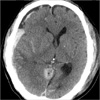
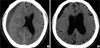
A 61-yr-old man was found unconsciousness following a high speed motor vehicle accident and he was immediately transferred to our institution. On admission, his Glasgow Coma Scale (GCS) score was 4 (E1V1M2). His pupils were fully dilated and non-reactive to light. The patient's laboratory results were within normal limits. The emergency computed tomography (CT) scan revealed a subdural hematoma with a maximal thickness of approximately 2.59 cm in the right fronto-temporo-parietal region with a severe midline shift of 2.42 cm, and this implied cerebral herniation (Fig. 1). The decision was made to treat him conservatively because of his poor clinical condition. With conservative management, an unexpected improvement in his neurologic status was noted within 14 hr of the trauma (GCS score; E1VtM3). A repeated CT scan showed a remarkable reduction in the amount of the hematoma and improvement of the midline shift from 2.42 cm to 1.10 cm (Fig. 2). It also demonstrated the new appearance of a high density lesion in the subdural space adjacent to the falx and tentorium, and a low-density area at the previous hematoma site, and especially over the left frontal convexity. An additional cerebral contusion was identified at the right temporal lobe. Thirty days after his admission, a CT scan revealed chronic SDH and an operation was done (Fig. 3A). The CT scan 7 days after the operation revealed brain atrophy and improvement of the effacement of the ipsilateral ventricle (Fig. 3B). The patient is currently able to obey simply commands and he still remains in a bed-ridden state.
DISCUSSION
ASDH is a sequel of severe injury and ASDH has a poor prognosis. Rapid spontaneous resolution of ASDH has rarely been reported because these patients usually undergo an operation on an emergency basis. The underlying pathophysiology is not well understood, but several possible mechanisms for the spontaneous resolution have been proposed. Two mechanisms are dilution of the hematoma by cerebrospinal fluid (CSF) due to tearing of the arachnoid membrane and this is followed by wash out (1-6) and compression of the hamatoma by acute brain swelling and this is followed by redistribution of the ASDH (5). The other theories are extracranial redistribution of the ASDH through a skull fracture (7), the presence of brain atrophy (3, 8-11), and development of subacute spinal SDH (12). However, the true mechanisms for spontaneous resolution of ASDH still remain unknown.
In our case, follow-up CT scan showed a dramatic reduction of the ASDH and a new high density region in the subdural space near the falx and tentorium. A low-density area at the anterior portion of the hematoma was also recognized, which implied involvement of CSF in the subdural space. We did not observe acute cerebral swelling. Sequential follow-up CT scanning revealed brain atrophy after evacuation of the chronic SDH. Cohen et al. (8) reported rapid spontaneous resolution of an acute subdural hematoma in a patient with cerebral atrophy and they described the AIDS related brain atrophy as the cause of this phenomenon. They postulated brain atrophy facilitated the redistribution and CSF washing out of the ASDH. Under these conditions, we suggest that the combined effect of a smaller brain and the increased amount of CSF in the subarachnoid space due to brain atrophy may favor both the redistribution and dilution of the ASDH. However, in our case, the amount of subdural blood was too large to be resolved spontaneously, although there was evidence of brain atrophy on the CT scan. Eventually, CT scan showed chronic SDH and an operation was done. Based on these observations, we think spontaneous resolution of a considerable amount of ASDH is possible, but there is an apparent limit of the amount of spontaneous resolution. Wu et al. (13), proposed that ASDHs might spontaneously resolve when their volume is less than 30 mL, when they are located in the fronto-temporal or tempro-parietal areas near the Sylvian fissure, when there is an isodence or low dense space between the hematoma and the inner wall of the skull on CT scan, and when they are associated with cerebral tumefaction and/or cerebral contusion and laceration in neurologically stable patients of a young age. Matsuyama et al. (4) reported complete disappearance of an ASDH with a thickness of 1.5 cm and they proposed that the absence of cerebral contusion is optimal for spontaneous resolution of ASDH. However, in our case, the thickness of the hematoma was 2.59 cm and there were cerebral contusions. Consequently, we suggest that these findings, including a huge amount of subdural blood (the thickness of hematoma above 2.5 cm) is an obstacle to complete spontaneous resolution of ASDH.
We experienced an unusual case in which the patient's neurologic condition was improved due to rapid spontaneous reduction of ASDH via conservative management, although the initial neurologic status was poor. However, in a real clinical setting, it is very difficult to distinguish which patients should be treated conservatively or surgically. Probably the most important parameter is the initial post-resusictation GCS score. The patient's age, their premorbid state and the arrival time to the hospital may also be considered. We selected conservative management in this case, but there is no confidence that we should apply the same management principles to other cases with a similar condition. Accordingly, it is mandatory to find clinical clues to help determine which patients should be treated with conservatively to manage a large amount of ASDH.
In conclusion, spontaneous rapid resolution/reduction of ASDH may occur in some patients, and even in patients with a large amount of ASDH. The mechanisms that are responsible for this are dilution by CSF and redistribution of the hematoma in patients with brain atrophy. We recommend that this treatment option should be used for carefully selected patients.

 XML Download