

 PDF
PDF ePub
ePub Citation
 Print
Print


INTRODUCTION
Adult-onset Still's disease (AOSD) is an inflammatory disease that presents with a variety of clinical symptoms, including fever, rash, arthritis, lymphadenopathy, and splenomegaly (1, 2). Fever is the dominant symptom, and infectious etiologies must be ruled out, as AOSD patients usually require immunosuppressive treatment (1, 2). Several complications associated with AOSD have been reported, such as cardiac dysfunction (3), aseptic meningitis (4), glomerulonephritis (5), thrombotic microangiopathy (6), and respiratory distress syndrome (7). In this report, we present a case of AOSD complicated with diffuse alveolar hemorrhage (DAH).
CASE REPORT
A 28-yr-old female presented with complaints of fever, fatigue, myalgia, skin rash, shoulder pain, and wrist swelling and pain. These symptoms had been present for a month, starting shortly after the patient had a sore throat for 5 days. Her body temperature was normal during the day, increasing to over 39℃ at night. A macular rash, concomitant with fever, was present on her trunk and arms. She was diagnosed as having pharyngitis in a peripheral hospital and was treated with several courses of antibiotics (amoxicillin-clavulanate, ciprofloxacin) without any diminishment of her symptoms. Her past medical history revealed fever, rash, and arthritis affecting her wrists 3- and 5-yr prior to the current episode, as well as use of nonsteroidal anti-inflammatory drugs, sulphasalasine, and a short course of oral corticosteroids for nearly 3 yr. In the past year, the patient had been symptom-free, and had discontinued her medications a year before the current admission.
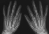
On physical examination at presentation, the patient's temperature was 39℃, and her heart rate was 100 bpm. She had painless cervical, axillary, and inguinal lymphadenopathy, and her spleen was palpable two centimeters below the left costal margin. Her pharynx was mildly erythematous. Musculoskeletal examination showed that her shoulders were painful when moved, and the motion of her wrists was also restricted due to pain. The rest of the examination was unremarkable. Routine laboratory investigation showed the following: erythrocyte sedimentation rate, 110 mm/hr; C-reactive protein, 347 mg/L; hemoglobin, 10.1 g/dL; platelets, 492,000/µL; total WBC count, 39,700/µL with a differential count of 90% neutrophils; ferritin level, 8,651 ng/mL. Cultures were negative for microorganisms, and antinuclear antigen and rheumatic factor were negative as well. An enzyme-linked immunosorbent assay test revealed negative IgM and positive IgG antibodies against the viral capsid antigen of the Epstein-Barr virus. An abdominopelvic ultrasonograph showed mild hepatomegaly and splenomegaly. An radiography of the patient's hands showed a typical presentation of bilateral carpal ankylosis (Fig. 1); the other radiography, including shoulders, feet, and sacroiliac joints, were normal. She was diagnosed with AOSD, and methylprednisolone (40 mg/day) was initiated.
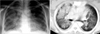
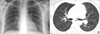
Four days after methylprednisolone was started, the patient developed progressive dyspnea and a cough. Complete blood count showed a decreased hemoglobin level (Hb: 6 g/dL) and leukocytosis (WBC: 18,700/µL). Her platelet count was normal (232,000/µL) and there was no fragmented red blood cells and spherocytes in the peripheral blood smear. Chest radiography revealed bilateral infiltration of her lungs and computed tomography showed bilateral consolidation, mediastinal lymphadenopathy, and pleural effusion (Fig. 2). Bronchoscopic examination showed hemorrhagic lavage, and further pathological examination revealed hemosiderin-containing macrophages. Repeated cultures, including culture of the bronchoalveolar lavage fluid, were negative. DAH secondary to AOSD was considered a likely diagnosis, and pulse methylprednisolone therapy was administered at a dose of 1,000 mg/day for 5 days. The patient's pulmonary symptoms improved dramatically after the first three doses of pulsed corticosteroids. After 5 days of pulsed methylprednisolone, therapy was maintained with 1 mg/kg methylprednisolone (40 mg/day) orally. Ten days after the pulse treatment, the chest radiography became normal (Fig. 3). Methotrexate was prescribed for refractory arthritis. No activation period was observed during the two-year follow-up period.
DISCUSSION
This case report describes a young female AOSD patient whose disease was complicated by DAH. Pulmonary involvement is well-known in AOSD and is seen in up to 53% of AOSD cases, with the most common pulmonary diseases being pleural effusion and transient pulmonary infiltrates (7). Life-threatening pulmonary complications, such as respiratory distress syndrome, are sometimes reported; however, to the best of our knowledge, DAH in AOSD has not been reported previously.
DAH is a medical emergency characterized by the accumulation of red blood cells in the alveolar spaces (8, 9). Hemoptysis, coughing, and progressive dyspnea are common initial symptoms, and the disease may progress to acute respiratory failure (8, 9). Hemoptysis is not a sine qua non symptom, and may be even absent in severe cases of DAH (8, 9). In cases of DAH, the chest radiograph is nonspecific, and most commonly shows new patchy or diffuse alveolar opacities (8, 9). Characteristic laboratory features of DAH are decreasing hematocrit and hemorrhagic bronchoalveolar lavage (8, 9). There have been many cases of DAH reported that include systemic vasculitis, collagen vascular diseases, drug-related factors, and infections (8, 9). Our patient was diagnosed as AOSD according to the Yamaguchi's classification, and she fulfilled all major and minor criteria (10). She had also experienced two other episodes in the past that included fever and arthritis, as well as having carpal ankylosis. As carpal and metacarpal ankylosis may be present in nearly 50% of chronic articular AOSD patients (1), we think that a history of fever and arthritis should also be included in the spectrum of symptoms that suggest an AOSD diagnosis. This patient was diagnosed as DAH due to her progressive dyspnea, cough, dramatically decreased hemoglobin levels, hemorrhagic bronchial fluid, and the hemosiderin-laden macrophages obtained from the bronchoalveolar lavage.
It is not known whether the association between AOSD and DAH is coincidental or whether there is some common pathogenic link. Several studies suggest that alterations in proinflammatory cytokine production play an important role in the pathogenesis of AOSD. Interleukin-18 (IL-18) appears to be the most important cytokine in AOSD pathogenesis, as it is overproduced in the acute phase of the disease and is believed to be the cytokine initiating the inflammatory cascade that includes interferon gamma, IL-6, and tumor necrosis factor alpha (11). On the other hand, a series of animal studies revealed that endotoxemia-related lung injury was associated with increased IL-18 levels in both blood and lung tissue (12). Furthermore, exogenous administration of IL-18 resulted in enhanced pulmonary microvascular leakage and neutrophil accumulation in the lung tissue of sham-operated mice (13). Based on these observations, we speculate that alterations in cytokines, especially increased IL-18 levels, might play an important role in the development of DAH in our patient.
In conclusion, DAH has not previously been reported in conjunction with AOSD, but this rare complication should be kept in mind when treating AOSD patients.
 XML Download