

 PDF
PDF ePub
ePub Citation
 Print
Print


INTRODUCTION
Epstein-Barr virus (EBV) is an oncogenic virus associated with various lymphoproliferative disorders (LPDs) (1-4). In immunocompetent hosts, the relative frequency of the occurrence, its clinical presentation, and EBV positivity of different histological subtypes of LPDs vary in different geographic areas, and this may be ascribed to genetic and environmental etiologic factors. In Western countries, Hodgkin's lymphoma (HD) and infectious mononucleosis are the most common EBV-associated diseases, whereas some EBV-associated LPDs, especially those associated with T lymphocytes or natural killer (NK) cells, are more prevalent in Asian and Latin American countries (5, 6).
T or NK cell LPDs associated with EBV have been differently named based on their clinical findings as well as the immunophenotype of proliferating cells. EBV-associated hemophagocytic lymphohistiocytosis (EBV-HLH) is an unusual syndrome characterized by fever, organomegaly, pancytopenia, and disseminated intravascular coagulation, and commonly occurs in children and adolescents (7). Chronic active EBV (CAEBV) infection has been reported mainly in Japan and is characterized by long-lasting infectious mononucleosis-like symptoms in children and young adults with no apparent immune deficiency (7). Fulminant T cell lymphoma following acute EBV infections, as described by Quintanilla-Martinez et al. (8), is characterized by hepatosplenomegaly-often without significant lymphadenopathy and by fever, liver failure, and hemophagocytic syndrome following recent viral-like upper respiratory illnesses. Aggressive NK cell leukemia (ANKL) is a neoplasm of NK cells that causes systemic illness and pursues an aggressive clinical course (9). Nasal-type NK/T cell lymphomas are distinct from other EBV-associated T or NK cell LPDs in their localization to the upper aerodigestive tract at the initial presentation, but once disseminated, they follow a similar fatal clinical course to other types of EBV-associated T or NK LPDs (10-12).
Previous studies on patients with EBV-associated T or NK LPDs mainly focused on the clinicopathological and virological aspects of a specific category of these diseases, but knowledge on the overall distribution among lymphoproliferative disorders and relationships between these diseases is lacking. This might be because such EBV-associated T or NK LPDs are uncommon, even in Asia.
Korea is an endemic area for EBV infection and shows a higher frequency of peripheral T or NK cell lymphomas than Western countries. There is a high prevalence of EBV infection in early childhood and nasal-type NK/T cell lymphomas are common, accounting for 8.7% of non-Hodgkin's lymphomas (NHL) in Korea (13). Thus, it is conceivable that many LPDs in Korea are associated pathogenetically with EBV and an analysis of their distribution may provide an insight into the relationships between each category of EBV-associated LPDs. Here, we analyzed the distribution and clinical findings for all patients with EBV-associated LPDs, with a special emphasis on the relationship among these diseases.
MATERIALS AND METHODS
Case selection
A total of 764 patients, including 67 children, were enrolled for the retrospective study. Among all lymphoproliferative disorders diagnosed in the Samsung Medical Center from 1994 to 2005, all patients with HD (n=52), T&NK cell lymphomas (n=226), and acute or chronic EBV-infections with or without subsequent development of LPD (n=21) were included for the study. To identify the frequency of EBV-positive B-cell NHL, 465 cases of B-NHLs for which paraffin blocks were available for study were retrieved from surgical pathology files. LPDs arising in immunocompromised hosts such as those receiving organ transplantation, or patients with congenital immune deficiency syndrome were excluded. Information for the EBV-encoded small nuclear RNAs (EBER) in situ hybridization study was available for all patients, either from previous studies performed during routine diagnostic work or from retrospective analysis. Clinical information, including age, sex, site of involvement, stage, and outcome, was obtained from the medical records.
Immunohistochemistry
Hematoxylin and eosin-stained slides were reviewed for all patients. Immunohistochemical analyses were performed on paraffin sections using monoclonal and polyclonal antibodies for the detection of lineage-specific or lineage-characteristic antigens. These included antibodies for CD3, CD4, CD8, CD20 (Novocastra, Newcastle upon Tyne, U.K.), CD10, CD15, CD21, CD23, CD30, cyclinD1, TIA-1, and granzyme B (DAKO, Glostrup, Denmark), and the anti-CD56 antibody (Novocastra). Sections of formalin-fixed tissue were stained with the avidin-biotin peroxidase procedure using diaminobenzidine as a chromogen. Immunohistochemical staining for LMP-1 and EBNA-2 (DAKO) was performed for selected EBER-positive tissues. Depending on the required protocol, the paraffin sections were pretreated in a microwave oven or with proteolytic enzymes for antigen retrieval.
Case definition
Acute EBV-associated hemophagocytic lymphohistiocytosis was defined as an acute EBV infection with fulminant manifestations such as persistent fever, severe hepatosplenomegaly, severe cytopenia, coagulopathy, hypertriglyceridemia, and/or hypofibrinogenemia, and histiocytic erythrophagocytosis in the bone marrow and secondary lymphoid organs (14). CAEBV infection was defined according to the following criteria suggested by Straus: 1) severe illness that had lasted more than 6 months and began as a primary EBV infection; 2) histological evidence of major organ involvement; 3) increased quantities of EBV in affected tissues detected by EBER in situ hybridization; and 4) no evidence of previous immunological abnormalities or other recent infections that might explain the observed condition (15). The lymphomas were classified according to the World Health Organization scheme (9).
EBER in situ hybridization
EBER in situ hybridization was performed for all specimens using paraffin-embedded tissue and fluorescein isothiocyanate-labeled oligonucleotide probe directed to EBV encoded RNA (EBER-1) (Novocastra). To identify cases with strong pathogenetic association with EBV in NHL, a positive reaction was defined only when more than 20% of the examined cells showed a nuclear signal. For HD, a positive case was defined when Hodgkin's cells showed a nuclear signal.
RESULTS
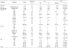
Demographic findings (Table 1)
The patients were all Korean, consisting of 299 women and 465 men, and had a mean age of 49 yr (range, 1-90). Histological subtypes included HD (n=52), B-cell NHL (n=465), T&NK cell lymphoma (n=226), acute EBV-HLH (n=7), CAEBV infection (n=9), and B-cell LPD arising in chronic EBV infection (n=5).
Malignant lymphomas (Table 1)
Histological type of EBV-positive malignant lymphoma (Table 1)
Among 743 cases of malignant lymphomas analyzed by in situ hybridization, 169 (23%) were EBER-positive. EBV was positive in 47% of T&NK cell lymphomas, 6.9% of B cell lymphomas, and 53.8% of HD. EBV was positive in almost all cases of NK/T cell lymphoma and aggressive NK cell leukemia. Compared with NK/T cell lymphoma or ANKL, less association was observed in other types of NHLs including diffuse large B cell lymphoma (DLBCL) (29/387, 7.5%), peripheral T cell lymphoma, unspecified (PTCL) (7/58, 12%), and Burkitt's lymphoma (3/26, 12%). In HD, EBER positivity was relatively high in those tumors with mixed cellularity (75%, 18/24), but low in lymphocyte-rich forms (40%, 2/5), and in nodular sclerosis (36%, 8/22).
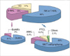
Among all EBV-positive T and NK cell type NHL cases, NK/T cell lymphoma was the most common subtype, accounting for 89 of 107 EBV-positive T or NK type NHL cases (83%). Other T or NK NHLs showed similar incidences: 8.4% of ANKL (9/107) and 6.5% of PTCL (7/107). EBV-positive B-NHL cases comprised with DLBCL (29/32, 91%) and Burkitt's lymphoma (3/32, 9%) (Fig. 1).
Age distribution of patients with EBV-positive malignant lymphoma
The ages of the patients with EBV-positive malignant lymphomas ranged from 5 to 88 yr with a median of 49. EBV-negative malignant lymphomas showed an age peak at the sixties whereas EBV-positive malignant lymphomas showed a broad peak from the forties to sixties.
The age distribution of patients with EBV-negative HD showed a major peak in the twenties, whereas the occurrence of EBV-positive HD showed a large peak from the forties to sixties and a smaller peak in the twenties. DLBCLs occurred in old patients, with an age peak in the sixties.
NK/T cell lymphoma showed the highest incidence in the forties. A few cases of EBV-associated PTCL and ANKL developed in the patients in the twenties, but the major peak was in the forties for ANKL and in the sixties for PTCL .
Clinicopathology of EBV-positive malignant lymphomas
The patients with EBV-positive NK/T cell lymphomas were 89 patients. Primary sites of presentation were the nasal cavity and nasopharynx in 62 patients and 27 in extranasal-sites. The clinicopathology of these patients has been reported previously (10, 11, 16). Most patients presented with local symptoms such as nasal obstruction, testicular enlargement, or abdominal pain, without any previous history suggesting chronic EBV infection or immune deficiency. Only one patient had a history of mosquito-bite hypersensitivity in childhood.
The nine patients with EBV-positive ANKL presented with acute syndromes of fever, pancytopenia, coagulopathy, and hepatosplenomegaly. These patients had no previous history suggestive of chronic EBV infection. Three of seven patients with EBV-positive PTCL showed a past history such as recurrent upper respiratory infection-like symptoms, hepatosplenomegaly, and increased liver enzymes with a duration shorter than 6 months, which suggested acute or subacute EBV infection. Other 3 patients with EBV-positive PTCL had a past or present illness of hepatitis C virus (HCV) infection, prostate cancer, or medication for rheumatoid arthritis.
There were 29 patients with EBV-positive DLBCL: 17 men and 12 women. Primary sites of presentation were lymph node in 12 patients and extranodal sites in 17 patients such as brain, peritoneum, and gastrointestinal tract. Most patients had no history suggestive of chronic EBV infection. Two patients had a concurrent illness such as hepatitis C and hepatitis B, and three patients had previous diseases such as tuberculosis, angioimmunoblastic T cell lymphoma, and HD.
Acute hemophagocytic lymphohistiocytosis and chronic EBV infection
The clinicopathology of patients with acute HLH, chronic active EBV infection associated with NK or T LPD, and B-LPD of various spectra arising in the background of chronic EBV infection was summarized in Tables 2, ,3, 4.
Among the patients with CAEBV infection, ANKL developed in two patients, hydroa-like T cell lymphoma in one, PTCL, unspecified in one, and monoclonal T-LPD in one patient.
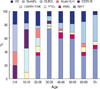
Common EBV-positive LPD type by age groups
In early childhood, Burkitt's lymphoma was the most common; in teenagers, chronic (active) EBV infection-associated LPD was the most common type. The incidence of NK/T cell lymphoma began to increase from the twenties and formed the major type of EBV-associated tumor throughout life. DLBCL increased from the fifties and tended to form the major type in the sixties and seventies (Fig. 2).
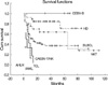
Comparison of the overall survival of patients with EBV-associated LPDs
Among 190 EBV-positive samples of LPDs, survival data were available for 177 patients (Fig. 3). The follow-up period ranged from a few days to 111.6 months, with a median of nine months and a mean of 19 months. Median patient survivals for each category of T or NK EBV-positive LPDs were 2 months for acute HLH, 1.6 months for ANKL, 5.5 months for PTCL, 8 months for T/NK LPD associated with CAEBV, and 13.2 months for NK/T cell lymphomas. In contrast to patients with T and NK LPD, those with B-lineage LPD and HD showed excellent prognosis; all five patients with B-LPD arising in chronic EBV infection were alive at follow-up. The median survival for patients with DLBCL and HD were 65 months and longer than 69.5 months, respectively.
DISCUSSION
Populations from different ethnic groups have variable susceptibility to EBV infection, as is demonstrated by geographical variations in the prevalence of EBV-related cancers. The relative incidence of T or NK/T cell lymphomas, especially EBV-associated nasal or nasal type NK/T cell lymphoma, is much higher in Asians than in Western populations (6, 13, 17). In Korea, T or NK/T cell lymphomas comprise 25% of NHLs (13), which is in line with reports from other Far Eastern countries. In the present study, T and NK cell lymphoproliferative diseases accounted for 64% of EBV-associated disease and 47.5% of T or NK cell lymphomas were associated with EBV. If we exclude those EBV-positive T and NK cell lymphomas, the incidence of NK and T cell lymphoma in Korea is similar to that of Western countries, accounting for 10-15% of NHLs (17), which again emphasizes the role of EBV in the pathogenesis of NK and T cell lymphomas in Asia.
B-cell tropism of EBV through the EBV-specific receptor CD21 (CR2), which is expressed on B cells and developing T cells but not on mature peripheral T cells, is well known (18). In individuals with a normal immune system, sustained T-cell infection by EBV occurs only rarely, raising the possibility that the infection of T lymphocytes and their subsequent unregulated growth is caused, at least in part, by a defect in immune surveillance (7). It has been suggested that a genetically determined susceptibility, possibly based on certain HLA types, results in an abnormal response to primary EBV infection in certain parts of Asia. A study of Chinese and natives of New Guinea has shown that this ethnic group has a high prevalence of HLA A11, a type that is associated with a mutation of EBNA-4 that abrogates cytotoxic T-cell recognition of EBV (19, 20). By contrast, A11 is rare among Europeans, in whom cytotoxic T cells recognizing the EBNA-4 peptide dominate the immune response to EBV. These variations in HLA phenotype may provide a basis for the higher frequency of EBV-positive tumors - including nasal T/NK cell lymphomas - among Asians. In addition, a recent study from Japan has shown that patients with nasal type EBV-associated NK/T cell lymphomas have a low frequency of the HLA-A*0201 allele, suggesting the importance of this allele in cytotoxic T lymphocyte responses (21).
CAEBV infection is a peculiar disease showing an abnormal immune response to primary EBV infection and characterized by recurrent infectious mononucleosis-like symptoms in childhood. Patients with CAEBV infections usually show clonal proliferation of T or NK cell and some of them develop overt NK cell or T cell lymphoma/leukemia in their teens and twenties (22). In the present study, ANKL and PTCL were mainly diseases of adults, but a few instances of EBV-associated PTCLs and ANKLs developed in patients in their twenties. Considering the natural course of chronic active EBV infection, these ANKL and PTCL occurring in young adults were probably transformed from chronic active EBV infections.
Patients with NK/T cell lymphomas, ANKL, and PTCL had major age peaks in their forties, forties, and sixties, respectively. It is not known what proportion of those EBV-associated diseases occurring in adults is a de novo disease unrelated with chronic (active) EBV infection. In Korea, the primary infection rate with EBV is 90% in those aged 7-9 yr and 100% in those between 10 and 15 yr (23). Almost all adult individuals are serologically positive. Therefore, EBV-associated diseases of adult onset seem to be derived from reactivation of chronic latent EBV infections or new EBV infection. In this study, we failed to find the past history suggesting chronic EBV infection from the medical record in most patients. Only one patient with a nasal-type NK/T cell lymphoma had mosquito-bite hypersensitivity when he was young. After childhood, he was relatively healthy without specific symptoms related to chronic EBV infection. Because mosquito-bite hypersensitivity is a cutaneous manifestation associated with latent EBV infection of the NK cells (7), this supports the idea that adult-onset nasal NK/T cell lymphoma may develop with a background of chronic latent EBV infection. Given the age that most patients develop NK/T cell lymphomas, transformation of EBV-infected NK cells into NK/T cell lymphoma cells may require a long latent period for the accumulation of genetic mutations sufficient for neoplastic transformation. Prospective epidemiologic studies are clearly needed to clarify the natural history of adult-onset EBV-associated malignancy.
In this study, some patients with adult-onset EBV-associated LPD showed associated illnesses that could have caused immunological dysfunctions provoking the reactivation of latent EBV infection and neoplastic transformation of EBV-infected cells. Four of six adult patients with acute EBV-HLH were associated with HCV hepatitis and chronic arthritis, or postpartum status. Three of seven patients with EBV-associated PTCL had a history of medication for rheumatoid arthritis, HCV hepatitis, and prostate carcinoma. Common association with HCV in these patients suggests an apparently ineffective antiviral T-cell response. Likewise, in the patients with DLBCL, 19 of the 29 patients were older than 60 yr and 5 of the 29 adult EBV-positive DLBCL patients had histories of HCV hepatitis, hepatitis B virus (HBV) hepatitis, tuberculosis, or HD, which suggests that decreased immunity in elderly patients may also contribute to the pathogenesis of adult EBV-positive DLBCL (24).
It is intriguing that the five patients with chronic EBV infection were associated with B-cell LPDs of various histological spectra. Three of four such patients presented with recurrent tonsillitis-like symptoms with monoclonal or oligoclonal B cell proliferation. Two of them expressed EBNA-2 as well as LMP-1, which indicates a type III latency of EBV infection usually identified in immunocompromised patients and which indicates an innate defect in the immune surveillance of EBV infection. The prognosis of those patients with B-LPD was excellent, as it was for those with EBV-associated large B cell lymphomas and for adult patients with HD. With additional genetic changes, these proliferating B cells in children with an immune defect to eradicate EBV may convert to HD or DLBCL, although the exact relationship between B-cell LPDs in children with chronic EBV infection and EBV-associated large B cell lymphomas or HD arising among adult patients remains to be clarified.
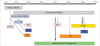
In summary, EBV infection in Koreans induced predominantly T or NK cell LPDs; among these, NK/T cell lymphomas were the most common. A high prevalence of EBV infection in early childhood associated with the failure of innate immune responses to eradicate the virus resulted in the development of EBV-associated LPD throughout life. As shown in Fig. 4, primary infections in early childhood may be complicated by the development of Burkitt's lymphoma and acute EBV-HLH as well as CAEBV infections; some of these transformed to ANKLs and PTCLs in young adults. In the middle-aged patients, some with chronic latent infections developed NK/T cell lymphomas and ANKL. In old patients, decreased immunity and environmental cofactors may provoke the development of PTCL and DLBCL.
 XML Download