

 PDF
PDF Citation
 Print
Print


It is with great interest that we read the case report entitled "Spontaneous vertebral reduction during the procedure of kyphoplasty in a patient with Kummell's disease" by Hur et al., which was published in the 2011 December issue of the Korean Journal of Pain [1]. First, we congratulate the authors on the good outcome achieved by the use of their good surgical skills in a patient on whom it was difficult to perform kyphoplasty. We also appreciate the fact that the authors explained the possible mechanism underlying this rare phenomenon through their extensive literature review. However, we would like to make some comments on their case report. We think that the title of the case report, i.e., "Spontaneous vertebral reduction during the procedure of kyphoplasty in a patient with Kummell's disease" is slightly erroneous. The medical dictionary definition of spontaneous is "developing without apparent external influence, force, cause, or treatment." However, the reduction of the fractured lumbar vertebrae in the authors' patient was caused by cannula insertion and did not occur spontaneously. Additionally, the prone position during the operation might have contributed to the reduction of the fractured vertebrae. Our comment is supported by the last paragraph on page 232 of the report that states the following: "At the moment when the left cannula was inserted into the vertebral body, air flowed into the vertebral body with a popping sound, and the level of the vertebral body recovered spontaneously (Fig. 3)." Although the phenomenon of height restoration after a popping sound is rare, it does sometimes occur during percutaneous kyphoplasty or vertebroplasty. We think that insertion of the cannula into the fracture line via the transpedicular approach can induce iatrogenic dynamic mobility of the fractured vertebra. Reduction is caused by cannula insertion and positional gravity. Therefore, we seriously recommend that the title of their case report be changed to "Cannula-induced vertebral reduction during kyphoplasty in a patient with Kummell's disease." In addition, we present the case of our patient, which is similar to that of the authors' patient.
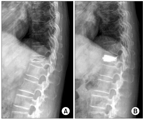
CASE REPORT
A 62-year-old woman presented with a 2-week history of severe pain in the lower back, which had started after she had slipped and fallen. Her bone mineral density (BMD) T-score was -1.5, and her visual analogue scale (VAS) score was 8.1. A radiograph showed severe vertebral collapse with a vertebral vacuum cleft in the T12 vertebral body (Fig. 1A). The T1-weighted magnetic resonance imaging (MRI) scans showed low signal intensity at T12, suggesting an acute compression fracture. Therefore, the patient underwent percutaneous vertebroplasty at L1 via the transpedicular approach. When we inserted the cannula into the fractured vertebral body, reduction of the fractured vertebrae developed. Postoperative radiography showed reduction of the collapsed T12 vertebral body (Fig. 1B).
 XML Download