

 PDF
PDF ePub
ePub Citation
 Print
Print


Abstract
Background:
Eosinophilia may be associated with various primary and reactive conditions. The incidence and the causes of eosinophilia might have been changed according to the changes in the incidence of diseases such as cancer, chronic degenerative diseases, etc. We have conducted a retrospective study to investigate the incidence and causes of eosinophilia.
Methods:
Eosinophilia and hypereosinophilia were defined when absolute eosinophil count was greater than 500/μL and 1,500/μL, respectively. Patient's clinical records were reviewed to find out the underlying clinical conditions responsible for causes of hypereosinophilia. Conventional chromosomal analysis, reverse transcriptase PCR and FISH for gene rearrangement were performed to check the presence of clonal eosinophilia.
Results:
Out of 41,137 patients who had a hematology profile performed, 5,019 (12.2%) and 373 patients (0.9%) were found to have eosinophilia and hypereosinophilia, respectively. Among patients with hypereosinophilia, 227 patients (60.9%) had identifiable and/or possible causes. The major causes of hypereosinophilia were malignancy (35.2%), allergy and skin diseases (18.1%), infectious diseases (15.4%), hepatobiliary diseases (7.5%), bone marrow clonal diseases (6.6%) and parasite infections (6.6%). We also found a rare case of FIP1L1-PDGFRα positive chronic eosinophilic leukemia combined with light chain multiple myeloma.
Conclusions:
We found a difference in the distribution of causes of hypereosinophilia in comparison with previous Korean studies, and the most common cause of hypereosinophilia in the current study was malignancy. A rare case of clonal eosinophilia (chronic eosinophilic leukemia) associated with multiple myeloma was confirmed using molecular studies.
REFERENCES
1.Brito-Babapulle F. Clonal eosinophilic disorders and the hypereosinophilic syndrome. Blood Rev. 1997. 1:129–45.

2.Wardlaw A. Eosinophils and their disorders. Lichtman MA, Beutler E, editors. Williams hematology. 7th ed.New York, NY: Mc Graw-Hill;2006. p. 863–78.
3.Brito-Babapulle F. The eosinophilias, including the idiopathic hypereosinophilic syndrome. Br J Haematol. 2003. 121:203–23.
4.Chung JP., Nam DK., Lee SJ., Lee EK., Hahn JS., Ko YW. A clinical study on eosinophilia; with a report of 5 cases of hypersoinophilic syndrome. Korean J Hematol. 1988. 23:127–37. (남 령, 강명석, 한진영, 이은엽, 김순호. 제대혈과 신생아 말초 혈의참고치.대한임상병리학회지 1988;23:127-37.).
5.Shin KS., Choi YM., Chae SA., Hyung SM. A clinical study of cause of Eosinophilia. Chungbuk Med J. 1996. 6:105–14. (신경섭, 최윤미, 채 수안, 형성민. 호산구증다증에대한임상적고찰. 충북의대학술지 1996;6:105-14.).
6.Shaffer LG, Tommerup N, editors. ISCN 2005: an international system for human cytogenetic nomenclature. 1st ed.Basel: Karger;2005.
7.Whitby LE, Britton CJC, editors. Disorder of the blood. Diagnosis: Pathology: Treatment: Technique. 9th ed.New York: Grune & Stratton Inc.;1968. p. 15.
8.Wintrobe MM, editor. Clinical hematology. 6th ed.Philadelphia: Lea & Febiger;1967. p. 260.
9.Orfanakis NG., Ostlund RE., Bishop CR., Athens JW. Normal blood leukocyte concentration values. Am J Clin Pathol. 1970. 53:647–51.
10.Zacharski LR., Elveback LR., Kinman JW. Leukocyte counts in healthy adults. Am J Clin Pathol. 1971. 56:148–50.
11.Teo CG., Singh M., Ting WC., Ho LC., Ong YW., Seet LC. Evaluation of the common conditions associated with eosinophilia. J Clin Pathol. 1985. 38:305–8.
12.Wykoff RF. Eosinophilia. South Med J. 1986. 79:608–12.
13.Tefferi A., Patnaik MM., Pardanani A. Eosinophilia: secondary, clonal and idiopathic. Br J Hematol. 2006. 133:468–92.
14.Leder K., Weller PF. Eosinophilia and helminthic infections. Baillieres Best Pract Res Clin Haematol. 2000. 13:301–17.
15.Slugaard A., Ascensao J., Zanjani E., Jacob HS. Pulmonary carcinoma with eosinophilia. Demonstration of a tumor-derived eosinophilopoietic factor. N Engl J Med. 1983. 309:778–81.
16.Anagnostopoulos GK., Sakorafas GH., Kostopoulos P., Margantinis G., Tsiakos S., Terpos E, et al. Disseminated colon cancer with severe peripheral blood eosinophilia and elevated serum levels of interleukine-2, interleukine-3, interleukine-5, and GM-CSF. J Surg Oncol. 2005. 89:273–5.
17.Dellon AL., Hume RB., Chretien PB. Letter: eosinophilia in bronchogenic carcinoma. N Engl J Med. 1974. 291:207–8.
18.Backenroth R., Spinowitz BS., Galler M., Golden RA., Rascoff JH., Charytan C. Comparison of eosinophilia in patients undergoing peritoneal dialysis and hemodialysis. Am J Kidney Dis. 1986. 8:186–91.
19.Bain B. Eosinophilic leukaemias and the idiopathic hypereosinophilic syndrome. Br J Haematol. 1996. 95:2–9.
20.Cools J., DeAngelo DJ., Gotlib J., Stover EH., Legare RD., Cortes J, et al. A tyrosine kinase created by fusion of the PDGFRA and FIP1L1 genes as a therapeutic target of imatinib in idiopathic hypereosinophilic syndrome. N Engl J Med. 2003. 348:1201–14.
21.Gotlib J. Molecular classification and pathogenesis of eosinophilic disorders: 2005 update. Acta Haematol. 2005. 114:7–25.
22.Bain BJ., Gilliland DG., Horny HP., Vardiman JW. Myeloid and lymphoid neoplasms with eosinophilia and abnormalities of PDGFRA, PDGFRB or FGFR1. Swerdlow S, Harris NL, editors. World Health Organization Classification of tumours. Pathology and genetics of tumours of haematopoietic and lymphoid tissues. 4th ed.Lyon: IARC Press;2008. p. 68–73.
23.Hardy WR., Anderson RE. The hypereosinophilic syndrome. Ann Intern Med. 1968. 68:1220–9.
24.Chusid MJ., Dale DC., West BC., Wolf SM. The hypereosinophilic syndrome; analysis of fourteen cases with review of the literature. Medicine. 1975. 54:1–27.
25.Bain BJ., Gilliland DG., Horny HP., Vardiman JW. Chronic eosinophilic leukaemia, not otherwise specified. Swerdlow S, Harris NL, editors. World Health Organization Classification of tumours. Pathology and genetics of tumours of haematopoietic and lymphoid tissues. 4th ed.Lyon: IARC Press;2008. p. 51–3.
26.Brigden M., Graydon C. Eosinophilia detected by automated blood cell counting in ambulatory North American outpatients. Incidence and clinical significance. Arch Pathol Lab Med. 1997. 121:963–7.
Fig. 1.
Morphology and the FIP1L1-PDGFRα rearrangement in a patient with clonal eosinophilia associated with multiple myeloma. (A) The BM aspiration smear shows diffuse plasma cell infiltration (27%) and increased number of mature eosinophils and eosinophil precursors (10%) (Wright stain, 400). (B) The BM section biopsy shows a hypercellular marrow with plasma cell clusters (H&E stain, ×100). (C) Abnormal nucleus showed one fusion signal (red arrow) and deleted orange signal (white arrow) which indicates the 4q12 using LSI 4q12 tricolor rearrangement probe (Vysis). (D) Reverse transcriptase-polymerase chain reaction (RT-PCR) analysis of RNA isolated from the bone marrow of the patient. The amplified band for FIP1L1-PDGFRα transcripts is shown on the right side of the figure. (E) The detection of FIP1L1-PDGFRα using PCR-direct sequencing analysis in the patient. The sequence trace (reverse chromatogram) result shows the break and fusion point (indicated by the arrow) in exon 12 of PDGFRα and exon 12 of FIP1L1 based on the RT-PCR results. Abbreviation: MW, molecular weight marker (Boehringer, Ingelheim, Germany).
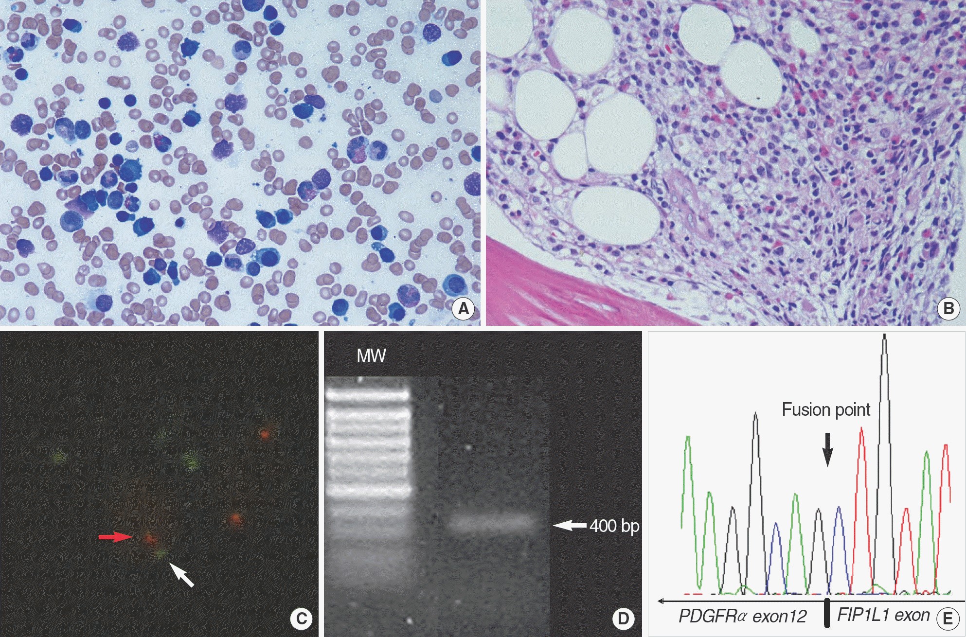
Table 1.
Identifiable and/or possible causes of eosinophilia
Table 2.
Comparison of mean eosinophil count in different clinical entities with moderate to severe eosinophilia
Table 3.
Comparison of incidence, number of patients, patient age, sex and etiologies in the previous studies and the present study
 XML Download