

 PDF
PDF ePub
ePub Citation
 Print
Print


Dear Editor,
Interactions of thyroid hormone autoantibodies (THAAs) with thyroxine (T4) or triiodothyronine (T3) are less common than interactions of antibodies to thyroglobulin, microsomal thyroid peroxidase, and thyroid-stimulating hormone (TSH) receptor. Nevertheless, these THAAs have been reported to cause spurious thyroid function test (TFT) results, indicating that TFT results do not always accurately reflect the clinical status of the patient and are not necessarily consistent [12].
The susceptibility to THAA interference depends particularly on whether the patient serum components are exposed to the tracers used (e.g., one-step vs two-step immunoassay, equilibrium dialysis, or ultrafiltration methods) and not the label used in the tracers. In two-step methods, all serum components are removed by a washing step before addition of the tracer [1]. Therefore, automated one-step immunoassays are known to be more susceptible to THAA interference [2]. We report the first case of the spurious elevation of free T4 in a Korean patient caused by circulating THAA.
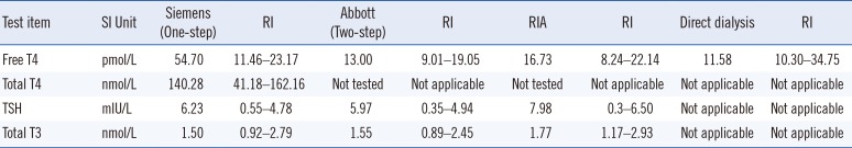
A 40-yr-old female presented with fatigue and edema spanning a three-month period. She had taken methimazole owing to elevated free T4 and TSH levels detected on a TFT performed in another hospital. For proper management, she was transferred to our hospital. The endocrinologist ordered a TFT from the Departments of Laboratory Medicine and Nuclear Medicine. Total T3, total T4, free T4, and TSH were measured with a one-step chemiluminescent immunoassay (CLIA) on the ADVIA Centaur XP system (Siemens Healthcare, Munich, Germany) and with an RIA. The serum concentrations of total T3 and free T4 were measured by using a total T3 RIA kit (Immunotech Inc., Praha, Czech Republic) and a free T4 RIA kit (Immunotech), respectively. Free T4 was quantitated with a two-step CLIA on an Architect i2000 system (Abbott Diagnostics, Santa Clara, CA, USA). The TFT results obtained from the Department of Laboratory Medicine were comparable with the results from the outside hospital (Table 1). By contrast, the results from the Department of Nuclear Medicine exhibited a decreased free T4 level and an unaltered total T3 level. The patient's clinical symptoms and elevated TSH level indicated hypothyroidism, and the patient was diagnosed as having Hashimoto's thyroiditis.
Potential causes of the spurious TFT results include nonspecific binding of endogenous circulating factors such as heterophilic antibodies, albumin variants found in familial dysalbuminemic hyperthyroxinemia (FDH), and THAA [3]. In the general population, heterophile antibodies could be detected within the range of 0.2–15% [1]. These antibodies seldom interfere with immunoassays (0.05–0.5%) [4]. Retesting using a heterophilic antibody blocking tube (Scantibodies, Santee, CA, USA) did not affect the patient's results. FDH is an autosomal dominant disease caused by ALB gene mutation increasing the affinity of albumin for T4 by approximately 60-fold. The prevalence is 1 in 10,000 people [3]. FDH is common causes of euthyroid hyperthyroxinemia with an increased circulating total T4, andprotein electrophoresis can be helpful to screen abnormal albumin [3].
While the one-step assay is vulnerable to THAAs, which directly compete with endogenous free T4, a two-step assay employing an intermediate washing step induces a non-competitive reaction that removes the unbound free T4 and interfering factors. When the patient's sample was retested by the two-step assay (Architect, Abbott Diagnostics) (Table 1), the free T4 level was not increased, which was concordant with the RIA results, whereas it was elevated in the one-step assay. This suggested that free T4 in the one-step assay was falsely elevated, implying the possibility of the presence of an anti-T4 antibody. The presence of T4 antibodies in the patient was identified by a radiobinding assay (Quest Diagnostics, San Juan Capistrano, CA, USA). Free T4 was measured by the reference method of direct equilibrium dialysis, which was compatible with the results measured by the RIA.
To our knowledge, this is the first Korean case of T4 autoantibodies influencing spurious TFT results. Two previous Korean case reports involved free T3 autoantibodies as a cause of spurious elevation of free T3 level [56]. These patients were also diagnosed as having Hashimoto's thyroiditis.
The frequency of THAAs is about 2% in the general population and about 30% in patients with autoimmune thyroid disease [7]. Despite its high prevalence, significant interference caused by THAA is relatively rare and depends on the qualitative characteristics of the autoantibody present (i.e., its affinity for the test reagents) [3].
Interference in immunoassays is encountered frequently and can adversely affect patient care. Laboratory staff should be aware of the possibility of the presence of THAA when TFT results do not reflect the clinical status. However, it is difficult to detect such interference proactively in a laboratory; thus, it is important to establish and maintain rapport with the clinicians treating the patient.
In conclusion, we report the first Korean case of spurious elevation of free T4 caused by circulating THAAs, indicating the clinical significance of THAA and the importance of cautious interpretation of TFT results that conflict with a patient's clinical presentation, especially in autoimmune diseases.
 XML Download