Journal List > Ann Lab Med > v.36(5) > 1091438

Lee, Kim, Lee, Park, Kim, Chang, Lee, Kim, Oh, and Song: Acute Myeloid Leukemia With MLL Rearrangement and CD4+/CD56+ Expression can be Misdiagnosed as Blastic Plasmacytoid Dendritic Cell Neoplasm: Two Case Reports
Dear Editor,
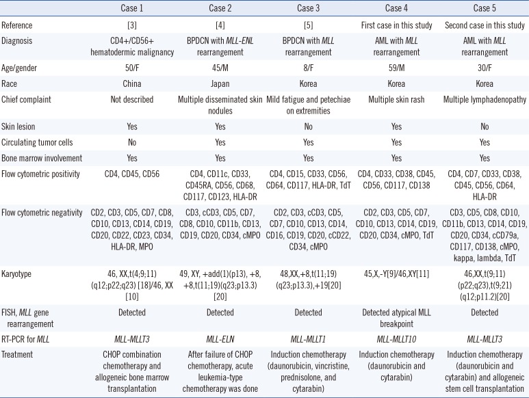
Blastic plasmacytoid dendritic cell neoplasm (BPDCN), characterized by co-expression of CD4 and CD56 without any other lineage-specific markers, is an aggressive tumor type that shows a high frequency of skin involvement and nodal or marrow infiltration with a propensity toward leukemic dissemination [1].This disease was formerly defined by the World Health Organization as blastic NK-cell lymphoma, and was later grouped with AML and related precursor neoplasms [2]. Here we report two AML cases with KMT2A (or MLL) rearrangements along with CD4+/CD56+ expression, which had the potential to be misdiagnosed as BPDCN.
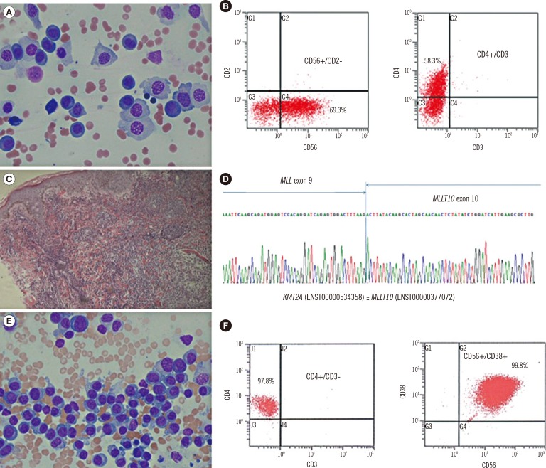
A 59-yr-old man presented with erythematous papules and vesicles on his whole body for one week. Complete blood counts showed anemia (Hb, 80 g/L), thrombocytopenia (62×109 platelets/L), and many circulating plasmoblast-like cells (65%). Bone marrow examination showed hypercellular marrow (90%) mostly composed of plasmoblast-like cells with coarse nuclear chromatin, distinct nucleoli, and abundant basophilic cytoplasm (Fig. 1A). Flow cytometry revealed that the neoplastic cells were positive for CD4, CD33, CD38, CD56, CD117, and CD138 (Fig. 1B), and negative for other myeloid or lymphoid markers (Table 1). Skin lesions showed diffuse infiltration of medium- to large-sized agranular blastic cells with CD4 and CD56 co-expression (Fig. 1C). Bone marrow and skin were negative for CD123 immunohistochemical stain. The chromosome study revealed 45,X,-Y[9]/46,XY[11] without 11q23 abnormalities. Multiplex, nested reverse transcription (RT)-PCR was performed for screening and detection of 28 chromosomal translocations using the HemaVision kit (DNA Technology, Research Park, Aarhus, Denmark), and the presence of MLL-MLLT10 rearrangement was demonstrated. It was confirmed by direct sequencing (Fig. 1D). Fluorescence in situ hybridization for MLL rearrangement revealed nuc ish(5'MLLx3,3'MLLx2)(5'MLL con 3'MLLx2)[170/200], suggesting the presence of an atypical MLL breakpoint. Although the lineage antigens expressed here were not specific to AML, he was diagnosed as having AML with MLL rearrangement in bone marrow and skin.
A 30-yr-old woman presented with multiple lymphadenopathy for three weeks. Laboratory analysis revealed anemia (Hb, 81 g/L), thrombocytopenia (82×109 platelets/L), and leukocytosis (16.77×10⁹ white blood cells/L) with circulating plasmacytoid cells (63%). Neither skin lesion nor monoclonal gammopathy was present. Bone marrow analysis showed hypercellular marrow (100%) with leukemic cells containing abundant basophilic cytoplasm (Fig. 1E). Flow cytometry revealed that the neoplastic cells were positive for CD4, CD7, CD33, CD38, CD56, and CD64, (Fig. 1F), and were negative for other markers (Table 1). The cytochemical stain for non-specific esterase in neoplastic cells was negative. Immunohistochemical stains of bone marrow biopsy specimens showed that the neoplastic cells were negative for CD123, CD138, kappa, and lambda. Lymph node biopsy analysis demonstrated that the neoplastic cells co-expressing CD4 and CD56 were negative for CD123. Cytogenetic analysis revealed 46,XX,t(9;11)(p22;q23),t(9;21)(q12;p11.2)[20]. The MLL-MLLT3 rearrangement was detected by RT-PCR. Although adequate lineage-specific markers were not observed, she was diagnosed as having an AML with MLL rearrangement in bone marrow and lymph nodes.
So far, three cases of CD4+/CD56+ hematologic malignancies with MLL rearrangements have been reported as rare cases of BPDCN with MLL rearrangement (Table 1). The leukemic cells reported in the literature expressed the myeloid and monocytic markers CD33, CD117, CD11c, CD15, or CD64, which were not specific enough to identify a specific lineage [345]. These three cases did not express highly specific plasmacytoid dendritic cell-associated antigens, such as CD123, TCL1, CD2AP, or CD303 (BDCA2); therefore, additional immunohistochemical studies were necessary at their diagnosis. Variable expression of CD4 or CD56 in adult AML cases with MLL rearrangement has been reported [6], and BPDCN is often confused with monocytic leukemias. Based on the limited information on immunophenotypes of these cases, MLL-related monocytic leukemia would be a reasonable diagnosis (Table 1).
This study emphasizes that the diagnosis of BPDCN should only be considered after a full investigation of plasmacytoid dendritic cell markers, ensuring there is no expression of myeloid or monocytic markers on the blasts [7]. Since hematologic neoplasms such as BPDCN, AML, extranodal NK/T cell lymphoma, nasal type, and mature T cell lymphomas with or without skin involvement may express CD56 with or without CD4, extensive immunohistochemical and genetic analyses are necessary before definitively diagnosing BPDCN or AML [89].
In conclusion, CD4+/CD56+ hematologic malignancies can be suspected to be BPDCN, and clinicians should conduct a full analysis including flow cytometry for adequate myeloid/monocytic markers, immunohistochemical stain for highly specific plasmacytoid dendritic cell-associated antigens, cytogenetic, and genetic studies to make an exact diagnosis and determine effective treatment.

Acknowledgments

This work was supported by the yearly clinical research grant from Pusan National University Yangsan Hospital.

Notes

Authors' Disclosures of Potential Conflicts of Interest: No potential conflicts of interest relevant to this article were reported.

References

1. Garnache-Ottou F, Feuillard J, Saas P. Plasmacytoid dendritic cell leukaemia/lymphoma: towards a well defined entity? Br J Haematol. 2007; 136:539–548. PMID: 17367408.
2. Swerdlow SH, Campo E. . WHO classification of tumours of haematopoietic and lymphoid tissues. France: IARC Press;2008. p. 145–147.
3. Leung R, Chow EE, Au WY, Chow C, Kwong YL, Lin SY, et al. CD4+/CD56+ hematologic malignancy with rearranged MLL gene. Hum Pathol. 2006; 37:247–249. PMID: 16426929.
4. Toya T, Nishimoto N, Koya J, Nakagawa M, Nakamura F, Kandabashi K, et al. The first case of blastic plasmacytoid dendritic cell neoplasm with MLL-ENL rearrangement. Leuk Res. 2012; 36:117–118. PMID: 21831428.
5. Yang N, Huh J, Chung WS, Cho MS, Ryu KH, Chung HS. KMT2A (MLL)-MLLT1 rearrangement in blastic plasmacytoid dendritic cell neoplasm. Cancer Genet. 2015; 208:464–467. PMID: 26164398.
6. Muñoz L, Nomdedéu JF, Villamor N, Guardia R, Colomer D, Ribera JM, et al. Acute myeloid leukemia with MLL rearrangements: clinicobiological features, prognostic impact and value of flow cytometry in the detection of residual leukemic cells. Leukemia. 2003; 17:76–82. PMID: 12529663.
7. Rush PS, Bennett DD, Yang DT. Hematopathology HP 15-5. ASCP case reports. 2015; HP 15-5:1–20.
8. Bekkenk MW, Jansen PM, Meijer CJ, Willemze R. CD56+ hematological neoplasms presenting in the skin: a retrospective analysis of 23 new cases and 130 cases from the literature. Ann Oncol. 2004; 15:1097–1108. PMID: 15205205.
9. Herling M, Jones D. CD4+/CD56+ hematodermic tumor: the features of an evolving entity and its relationship to dendritic cells. Am J Clin Pathol. 2007; 127:687–700. PMID: 17439829.
Fig. 1

Morphological features, flow cytometric analysis, immunohistochemical stain, and genetic study of the two cases of CD4+/CD56+AML. (A) Plasmoblast-like neoplastic cells in the first case (Wright Giemsa stain, 400×, Bone marrow); (B) Immunophenotyping features with CD56 and CD4 coexpression in the first case; (C) Skin biopsy showing diffuse infiltration of medium- to large-sized agranular blastic cells into the dermis in the first case (Hematoxylin and Eosin stain, 100×, Skin lesion); (D) The MLL-MLLT10 rearrangement confirmed by direct sequencing in the first case; (E) Plasmacytoid cells in the second case (Wright Giemsa stain; ×400, Bone marrow); and (F) The immunophenotyping features with CD4 and CD56 coexpression in the second case.

alm-36-494-g001
Table 1

The clinicopathologic characteristics of CD4+/CD56+ hematologic malignancies carrying the MLL rearrangement

alm-36-494-i001
Case 1 Case 2 Case 3 Case 4 Case 5
Reference [3] [4] [5] First case in this study Second case in this study
Diagnosis CD4+/CD56+ hematodermic malignancy BPDCN with MLL-ENL rearrangement BPDCN with MLL rearrangement AML with MLL rearrangement AML with MLL rearrangement
Age/gender 50/F 45/M 8/F 59/M 30/F
Race China Japan Korea Korea Korea
Chief complaint Not described Multiple disseminated skin nodules Mild fatigue and petechiae on extremities Multiple skin rash Multiple lymphadenopathy
Skin lesion Yes Yes No Yes No
Circulating tumor cells No Yes Yes Yes Yes
Bone marrow involvement Yes Yes Yes Yes Yes
Flow cytometric positivity CD4, CD45, CD56 CD4, CD11c, CD33, CD45RA, CD56, CD68, CD117, CD123, HLA-DR CD4, CD15, CD33, CD56, CD64, CD117, HLA-DR, TdT CD4, CD33, CD38, CD45, CD56, CD117, CD138 CD4, CD7, CD33, CD38, CD45, CD56, CD64,
Flow cytometric negativity CD2, CD3, CD5, CD7, CD8, CD10, CD13, CD14, CD19, CD20, CD22, CD23, CD34, HLA-DR, MPO CD3, cCD3, CD5, CD7, CD8, CD10, CD11b, CD13, CD19, CD20, CD34, cMPO CD2, CD3, cCD3, CD5, CD7, CD10, CD13, CD14, CD16, CD19, CD20, cCD22, CD34, cMPO CD2, CD3, CD5, CD7, CD10, CD13, CD14, CD19, CD20, CD34, cMPO, TdT CD3, CD5, CD8, CD10, CD11b, CD13, CD14, CD19, CD20, CD34, cCD79a, CD117, CD138, cMPO, kappa, lambda, TdT
Karyotype 46, XX,t(4;9;11)(q12;p22;q23) [18]/46, XX [10] 49, XY, +add(1)(p13), +8, +8,t(11;19)(q23;p13.3)[20] 48,XX,+8,t(11;19)(q23;p13.3),+19[20] 45,X,-Y[9]/46,XY[11] 46,XX,t(9;11)(p22;q23),t(9;21)(q12;p11.2)[20]
FISH, MLL gene rearrangement Detected Detected Detected Detected atypical MLL breakpoint Detected
RT-PCR for MLL MLL-MLLT3 MLL-ELN MLL-MLLT1 MLL-MLLT10 MLL-MLLT3
Treatment CHOP combination chemotherapy and allogeneic bone marrow transplantation After failure of CHOP chemotherapy, acute leukemia-type chemotherapy was done Induction chemotherapy (daunorubicin, vincristine, prednisolone, and cytarabin) Induction chemotherapy (daunorubicin and cytarabin) Induction chemotherapy (daunorubicin and cytarabin) and allogeneic stem cell transplantation

Abbreviations: BPDCN, blastic plasmacytoid dendritic cell neoplasm; RT-PCR, reverse transcription-PCR; cMPO, cytoplasmic myeloperoxidase; TdT, terminal deoxynucleotidyl transferase; CHOP, cyclophosphamide, vincristine, epirubicin, and prednisolone.

TOOLS
Similar articles