

 PDF
PDF ePub
ePub Citation
 Print
Print


INTRODUCTION
Helicobacter cinaedi is a microaerophilic, motile, gram-negative spiral bacillus. This bacterium is part of the normal flora inhabiting the lower gastrointestinal tract of hamsters, and it has been suggested that pet hamsters serve as a reservoir for transmission to humans [1]. H. cinaedi was first isolated from the rectal swabs of homosexual men in 1984 [2], and thereafter, cases of bacteremia, gastroenteritis, and cellulitis caused by H. cinaedi have been reported. According to a study in Japan, the H. cinaedi-positive rate was 0.036% for blood cultures and 0.22% for positive blood cultures [3], indicating that this organism is not extremely rare. H. cinaedi is a typically opportunistic pathogen in immunocompromised patients, but bacteremia in immunocompetent hosts has also been reported [4]. In this study, we report the recovery and identification of H. cinaedi from the blood of a postsplenectomy patient for the first time in Korea.
CASE REPORT
A 71-yr-old man was admitted to the emergency room on November 9, 2011, because of dyspnea that developed 1 day ago. He had undergone splenectomy 3 yr ago because of immune hemolytic anemia. Subsequently, he developed aplastic anemia, and antithymocyte globulin and cyclosporine were administered until 1 yr ago. He had undergone minimally invasive direct coronary artery bypass surgery the previous December to treat angina pectoris.
On admission, his body temperature, pulse rate, respiration rate, and blood pressure were 36.4℃, 113/min, 25/min, and 133/76 mmHg, respectively. A complete blood cell count in the emergency room revealed a hemoglobin level of 9.3 g/dL, a white blood cell count of 1,500/µL with 25.0% neutrophils, and a platelet count of 11,000/µL. His C-reactive protein (CRP) level was elevated to 6.01 mg/dL at the time of admission. Chest plain radiography revealed pulmonary edema and bilateral pleural effusion. He was treated empirically with piperacillin/tazobactam for 9 days, although 3 sets of blood cultures using BACTEC Plus Aerobic/F and BACTEC Lytic/10 Anaerobic/F culture bottles (Becton-Dickinson, Franklin Lakes, NJ, USA) were all negative after a 5-day incubation. His absolute neutrophil count (ANC) recovered to 860/µL on hospital day (HD) 10 and 1,270/µL on HD 21. He developed a fever of 38.2℃ and his CRP level increased to 13.44 mg/dL on HD 21. Gram-negative spiral bacteria were recovered from the aerobic vials of all 3 sets of blood cultures after a 48 hr incubation (Fig. 1).
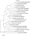
The organism did not grow on blood agar plates (BAPs) or MacConkey agar plates under 5% CO2, but it formed tiny translucent colonies that were 0.5 mm in diameter on chocolate agar plates after 3 days of incubation under microaerophilic conditions. This organism was able to grow at 35℃ and 42℃ but not at 25℃. It was positive for catalase and oxidase but negative for urease and indoxyl acetate hydrolysis. The isolate was first identified as Campylobacter spp. based on its growth characteristics and biochemical test results on HD 26. Because its poor growth on BAPs, MacConkey agar plates, and chocolate agar plates and negative results for the indoxyl acetate hydrolysis test were not compatible with its classification as Campylobacter spp., 16S rRNA gene sequencing was performed for further identification. Using primers amplifying 9-806 bp (8FPL 5'-AGT TTG ATC CTG GCT CAG-3', 806R 5'-GGA CTA CCA GGG TAT CTA AT-3') and 515-1,390 bp (515FPL 5'-TGC CAG CAG CCG CGG TAA-3', 13B 5'-AGG CCC GGG AAC GTA TTC AC-3'), PCR was performed according to previously published methods [5]. The purified PCR products were directly sequenced using the Big-Dye Terminator v3.1 Cycle Sequencing kit (Applied Biosystems, Foster City, CA, USA). According to a search of the Basic Local Alignment Search Tool (BLAST) database (http://www.ncbi.nlm.nih.gov/blast/), the sequence of this isolate exhibited 99.8% similarity (1,315 of 1,317 bp) with that of H. cinaedi CCUG 18818T (GenBank accession no. ABQT01000054). As secondary matches, the sequence of the isolate exhibited 98.5% and 97.9% with those of Helicobacter bilis Hb1T (GenBank accession no. U18766) and Helicobacter canis NCTC 12739T (GenBank accession no. L13464), respectively. Phylogenetic analysis was accomplished by using the MEGA 4.01, Molecular Evolutionary Genetic Analysis web-based software package [6]. Phylogenetic tree (Fig. 2) was constructed with MEGA4 using the Maximum Composite Likelihood method, and revealed this isolate to be H. cinaedi. The results of biochemical tests were also consistent with this identification.
The organism grew as a swarming thin film on brucella agar containing 7% sheep blood. Because this organism did not grow on Mueller-Hinton agar containing 5% sheep blood, antimicrobial susceptibility tests using disk diffusion and E test methods were performed with brucella agar containing 7% sheep blood [7]. Otherwise, we followed the testing conditions for Campylobacter jejuni/coli [8]. E tests for metronidazole and tetracycline revealed minimal inhibitory concentrations of 0.64 µg/mL, each. Using the disc diffusion method, the zones of inhibition of amoxicillin (2 µg), clindamycin (2 µg), erythromycin (15 µg), clarithromycin (2 µg), ciprofloxacin (5 µg), and trimethoprim-sulfamethoxazole (1.25 µg) were all found to be 6 mm in diameter, suggestive of resistance to these antimicrobials. The zones of inhibition of penicillin (10 µg), ampicillin (10 µg), rifampin (5 µg), amoxicillin/clavulanic acid (30 µg), and ceftriaxone (30 µg) were 16, 26, 52, 40, and 50 mm in diameter, respectively.
Piperacillin/tazobactam and levofloxacin were empirically administered for 11 days. The patient's body temperature normalized on HD 22, and his CRP level decreased to 2.74 mg/dL on HD 30. On HD 31, he was discharged without any symptoms or signs.
DISCUSSION
We isolated H. cinaedi from the blood cultures of an asplenic patient. The bacterium was identified using 16S rRNA gene sequencing. In this case, H. cinaedi was recovered from all 3 sets of blood cultures, and it was the only microorganism cultured from this febrile patient. Therefore, it was a true pathogen. Bacteremia is a common manifestation of H. cinaedi infection. It is believed that flagellar movement aids the adherence of H. cinaedi to the mucosal epithelium [9], and by producing a cytolethal distending toxin, it damages epithelial cells and invades blood vessels [10].
H. cinaedi bacteremia occurs primarily in immunocompromised hosts, particularly in men infected with HIV. Less commonly, infection may be observed in patients with alcoholism, diabetes, or malignancy, and occasionally in patients with no recognized defect in host defense [11]. The patient in this case study had previously undergone splenectomy. Although he had a history of aplastic anemia, he did not receive immune suppressive treatments in the previous year before hospitalization, and his ANC was 1,270/µL at the time of bacteremia onset. Therefore, splenectomy appears to have been a major risk factor for this bacteremia. There are a few reports of Campylobacter bacteremia in asplenic patients [12, 13]. Considering the common inhabitance of enterohepatic Helicobacter and Campylobacter spp., splenectomy would appear to be a risk factor for H. cinaedi bacteremia. There are no previous reports of H. cinaedi bacteremia in asplenic patients. Because the spleen plays a fundamental role in bacterial clearance, asplenia is a well-known risk factor for bacteremia. The most common bacteria that cause serious infections in asplenic patients are encapsulated organisms such as Streptococcus pneumoniae, Haemophilus influenzae type b, and Neisseria meningitidis [14]. Gram-negative rods are also involved in the development of infections in asplenic patients, and these infections are often fatal because of lipopolysaccharide-mediated inflammation [15]. It is also likely that splenectomized patients are immunocompromised because of multiple blood transfusions, chronic viral infections, iron overload, and diabetes mellitus [13].
The infectious organism in this patient was first identified as Campylobacter spp. H. cinaedi was originally identified as Campylobacter-like organism-1 but was transferred to the genus Helicobacter in 1989 [16]. Because of its helical shape and oxidase- and catalase-positive characteristics, it can be easily confused with Campylobacter spp. [17]. Because H. cinaedi is also fastidious and frequently exhibits unusual phenotypic profiles, microbiological diagnosis is difficult [3]. According to a study of 26 human isolates of H. cinaedi, the rates of catalase, nitrate, and indoxyl acetate positivity among the strains were 79%, 100%, and 6%, respectively. In addition, resistance to nalidixic acid and cephalothin was observed in 29% and 81% of strains, respectively, and growth at 42℃ was variable [18]. There is no single biochemical test to distinguish Helicobacter spp. from Campylobacter spp. [19]. A strain of H. cinaedi exhibiting resistance to both nalidixic acid and cephalothin may be difficult to differentiate from Campylobacter lari; however, H. cinaedi will not grow on MacConkey agar or in the presence of 1.5% NaCl [18]. In this study, 1,383-bp sequences of 16S rRNA were sufficient for identifying the organism at the species level [20]. It has also been reported that whole-cell protein or fatty acid analysis, or restriction profile analysis of the 23S rRNA gene, should be considered for identifying H. cinaedi [21]. However, these tests are not readily available in most clinical laboratories. Therefore, 16S rRNA sequencing is the most useful method for identifying H. cinaedi.
H. cinaedi is present in the normal intestinal flora of hamsters and rhesus monkeys [1, 22]. The clinical significance of H. cinaedi in human stool cultures is not clear [9]. Prior contact with animals has been reported in H. cinaedi-infected patients, suggesting that contact with carrier animals is a source of acquisition [22-24]. As a mode of transmission, the fecal-oral route was suspected, and there have been a few reports of nosocomial transmission [9, 25]. Stool cultures were not examined for H. cinaedi for this patient. Colitis is one of the primary clinical features of H. cinaedi infection [11]. In our patient, fever was the only prominent feature. Previous studies also revealed that most patients with H. cinaedi bacteremia have fever, but diarrhea is observed less frequently [3, 9, 23].
The infectious organism was resistant to macrolides and ciprofloxacin. In contrast to Campylobacter jejuni, H. cinaedi is not susceptible to erythromycin, and it exhibits variable susceptibility to fluoroquinolone [11, 23]. Because ciprofloxacin is commonly used for the empirical treatment of C. jejuni infection and other causes of bacterial enteritis, it is important to differentiate H. cinaedi from Campylobacter spp. This isolate was susceptible to most β-lactams, and therefore, piperacillin/tazobactam appeared to be sufficiently effective to eradicate it. Although treatments for H. cinaedi remain to be standardized, a previous report indicated that treatment with a penicillin, tetracycline, or aminoglycoside for 2-6 weeks was effective [23].
This is the first reported case of H. cinaedi bacteremia in Korea. We conclude that 16S rRNA gene sequencing is required to identify Campylobacter-like organisms at the species level. Further, asplenia appears to be a risk factor for H. cinaedi bacteremia.

 XML Download