

 PDF
PDF ePub
ePub Citation
 Print
Print


INTRODUCTION
The complications of preterm birth (PTB) cause approximately 70% of neonatal deaths and nearly half of all long-term neurological morbidity [1, 2]. PTB could be categorized by its clinical presentation: spontaneous preterm labor (PTL) leading to spontaneous PTB (S-PTB), preterm premature rupture of the membranes (PPROM), and medically induced PTB (M-PTB) due to maternal or fetal complications [3]. However, not all contributory causes of S-PTB have been identified, and the healthcare system is unable to target and manage relevant risk factors appropriately [4].
Intrauterine infection has been proposed as one of the most important risk factors for complications in pregnancy, such as premature rupture of membrane, PTL, PTB, and perinatal infections. Microorganisms may gain access to the amniotic cavity and the fetus through the following pathways: 1) ascending from the vagina and the cervix; 2) hematogenous dissemination through the placenta (transplacental infection); 3) retrograde seeding from the peritoneal cavity through the fallopian tubes; and 4) accidental introduction at the time of invasive procedures such as amniocentesis, percutaneous fetal blood sampling, chorionic villous sampling, or shunting [5]. Most studies on detection of infection in patients who underwent PTL and preterm delivery have focused on microbial invasion of the amniotic cavity, which is normally sterile. Therefore, the isolation of any microorganism from the amniotic fluid constitutes evidence of microbial invasion. The most common pathway of intrauterine infection is the ascending route [6].
Although colonization of the maternal genital tract by specific organisms has been inconsistently associated with S-PTB and/or PPROM, some infections have been consistently associated with preterm delivery [7]. It has been hypothesized that screening for and treatment of common vaginal infections would reduce the rate of PTB among affected women [8].
The microorganisms associated with PTL without preterm delivery (PTL-WO), S-PTB, and PPROM are as follows: Treponema pallidum, Neisseria gonorrhoeae, group B streptococcus (GBS), Ureaplasma urealyticum, Mycoplasma hominis, Chlamydia trachomatis, Trichomonas vaginalis, Gardnerella vaginalis, Bacteroides spp., and peptostreptococci [7, 9]. However, it is difficult to assess the extent of the causal relationship between infection and S-PTB because the maternal genital colonization rates by microorganisms differ according to race, gestational age, geographical variation, and investigators [2, 7, 9]. Moreover, it is not known how and when bacteria invade the uterus and whether additional (as yet undocumented) infections by viruses, protozoa, or bacteria other than those mentioned above are involved in PTB [9].
The purpose of this study was to determine the prevalence and antimicrobial susceptibilities of vaginal microorganisms isolated from Korean women who experienced PTL-WO and S-PTB. Finally, we sought to identify the microorganisms that were potential risk factors for PTB.
METHODS
1. Study population
The study population consisted of 126 pregnant women who experienced PTL and a control group of 91 pregnant women, with no history of PTL or preterm delivery and who underwent routine prenatal care, admitted to the Wonju Christian Hospital between August 2008 and July 2009. The patients were divided into the following 3 groups according to the type of complications during pregnancy: 1) PTL-WO (N=26), 2) S-PTB (N=69), and 3) M-PTB (N=31). M-PTB was defined as PTB with risk factors such as spontaneous rupture of membranes, anomalies of conception, malformations of the fetus, overdistended uterus, hydramnios, multiple gestations, fetal death, cervical incompetency, uterine anomalies, retained intrauterine device, serious maternal disease, pre-eclampsia, gestational diabetes mellitus, gestational hypertension, and active systemic infection. This study protocol was approved by the Institutional Review Board of Yonsei University Wonju College of Medicine.
2. Genital microorganisms
1) Culture and antimicrobial susceptibility test for GBS
Swab specimens from the vagina, anorectum, and urethral orifice of pregnant women were placed in selective Todd-Hewitt broth (S-THB) and new Granada plate and tube medium made in the laboratory [10] and in new Granada medium purchased from bioMérieux (Marcy-l'Etoile, France) for 18-24 hr in 5% CO2 atmosphere at 35℃. The colonies exhibiting orange coloration on new Granada tube or plate medium and/or exhibiting growth turbidity in S-THB were subcultured on 5% sheep blood agar and were identified using Streptex group B Streptococcus reagent (Wellcome Diagnostics, Dartford, England). Susceptibility to clindamycin, erythromycin, chloramphenicol, levofloxacin, penicillin, ceftriaxone, and vancomycin was tested using the MicroScan® MICroSTREP plus panel (Siemens Healthcare Diagnostics, Sacramento, CA, USA). The panel was inoculated using the Renok hydrator/inoculator, which delivered 115 µL of Mueller-Hinton broth with 3% lysed horse blood to each well. After inoculation with 0.5 McFarland standard bacterial suspension, the panels were incubated at 35℃ in ambient air for 20 to 24 hr and read using the MicroScan® WalkAway System (Siemens Healthcare Diagnostics).
2) Culture and antimicrobial susceptibility test for M. hominis and U. urealyticum
Vaginal swab samples for Mycoplasma IST 2 (bioMérieux) test were obtained from each pregnant woman; the test was performed according to the manufacturer's instructions. The Mycoplasma IST 2 test contains strips that indicate the presence or absence of M. hominis and U. urealyticum, and provide additional information on antibiotic susceptibility to tetracycline, doxycycline, erythromycin, clarithromycin, azithromycin, josamycin, ciprofloxacin, ofloxacin, and pristinamycin. One strip was placed directly into R1 tubes (transport medium) and subsequently delivered to the clinical microbiology laboratory for the identification of both M. hominis and U. urealyticum and for the determination of antimicrobial susceptibility. Swabs in the R1 transport medium were processed according to the manufacturer's instructions. They were mixed rapidly by vortexing, and then 3 mL of R1 was used to rehydrate the lyophilized growth medium R2 (provided in the Mycoplasma IST-2 kit). A Mycoplasma IST strip, consisting of 22 wells, was then inoculated with the rehydrated R2 growth medium (55 µL per well, overlaid with 2 drops of mineral oil). From the R2 positive tube, 0.1 mL was also inoculated onto A7 Mycoplasma agar plates (BioMérieux) and incubated at 37℃ in an atmosphere of 5% CO2 to evaluate characteristic colony morphology. All media and the inoculated strip were incubated at 37℃ in a CO2 incubator and monitored for color changes. The results were interpreted after 24 and 48 hr of incubation [11]. Wells 1-5 provided information on the presence or absence of M. hominis and U. urealyticum, with an estimate of the density of each organism (above, below, or equal to 104 colony-forming units [CFUs]). The antimicrobial susceptibility test was performed when the colony count for each organism was ≥104 CFU/mL.
3) Multiplex PCR for genital microorganisms
Samples for multiplex PCR were obtained from the posterior vaginal fornix by swabbing with a cytobrush. The swab was rinsed in a transport tube containing PCR buffer (phosphate-buffered saline solution, pH 7.4, without calcium chloride and magnesium chloride; GibcoBRL, Grand Island, NY, USA). T. vaginalis, M. hominis, Mycoplasma genitalium, U. urealyticum, C. trachomatis, N. gonorrhoeae, T. pallidum, herpes simplex virus (HSV) I, and HSV II were simultaneously detected using the Seeplex® STD6B ACE Detection kit (Seegene, Seoul, Korea). The swabbed sample was briefly vortexed and then centrifuged at 3,000 rpm for 20 min. After centrifugation, the mixture of sample pellet, proteinase K (Boehringer Mannheim, Mannheim, Germany), and buffer solution (10 mM Tric HCl, pH 8.3, 50 mM KCl, 0.1 mg/mL gelatin, 0.45% NP40, and 0.45% Tween 20) was incubated at 56℃ for more than 3 hr. The pellet was resuspended with phenol-chloroform, sodium acetate, and ethanol and incubated on ice for 30 min. Then, the DNA was extracted by centrifugation at room temperature for 5 min at 14,000 rpm, and the final pellet was frozen at -20℃. The extracted DNA was washed with 70% ethanol and resuspended in sterile distilled water before using it for multiplex PCR. Multiplex PCR was performed using the method described by Kim et al. [12].
4) Wet mount, Gram staining, and routine culture of vaginal specimens
Vaginal samples obtained using sterile cotton-tipped swabs were sent to the laboratory in transport medium. In the laboratory, the samples were aliquoted into 2 separate tubes: one for direct wet-mount microscopic examination and the other for Gram staining and subsequent inoculation onto blood and MacConkey agar plates. Examination of wet preparations was performed to rule out the presence of T. vaginalis and to detect the presence of yeasts. Gram staining was used to detect the presence of yeasts, inflammatory cells, and possible pathogenic microorganisms and to diagnose bacterial vaginosis (BV) by observing the "clue cells".
3. Statistical analysis
Statistical analysis was performed using the chi-square or Fisher's exact test with PASW Statistics 18 (SPSS Inc., Chicago, IL, USA). P values <0.05 were considered statistically significant. Relative risk ratio analysis was performed to evaluate if M. hominis, U. urealyticum, and GBS were independent risk factors for the development of S-PTB from PTL-WO.
RESULTS
1. The prevalence of genital microorganisms in different types of pregnancy complications
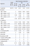
The colonization rates of GBS in the PTL-WO, S-PTB, M-PTB, and control groups were 3.8%, 8.7%, 9.7%, and 17.6%, respectively. Although the colonization rate of GBS in the S-PTB group was more than 2 times that in the PTL-WO group, there was no statistically significant difference between the 2 groups (relative risk ratio [RR], 2.26; 95% confidence interval [CI], 0.29-17.89; P>0.05). The colonization rate of GBS in the control group was much higher than that in the patient groups, although there was no statistically significant difference among the groups. The detection rates of M. hominis in the PTL-WO, S-PTB, and M-PTB groups were 3.8%, 17.3%, and 9.7%, respectively. The prevalence of M. hominis in the S-PTB group was higher than that in the PTL-WO group; however, there was no statistically significant difference between the 2 groups (RR, 4.52; 95% CI, 0.62-33.06; P=0.105). The detection rates of M. hominis by PCR and culture were as follows: 3.8% vs. 3.8% in the PTL-WO group, 14.4% vs. 5.7% in the S-PTB group, and 9.7% vs. 0% in the M-PTB group. The detection rates of U. urealyticum in the PTL-WO, S-PTB, and M-PTB groups were 53.8%, 60.9%, and 74.2%, respectively. The prevalence of U. urealyticum in the S-PTB group was higher than that in the PTL-WO group; however, there was no statistically significant difference between the 2 groups (RR, 1.13; 95% CI, 0.76-1.69; P>0.05). The detection rates of U. urealyticum by PCR and culture were as follows: 19.2% vs. 42.3% in the PTL-WO group, 17.4% vs. 58.0% in the S-PTB group, and 13.0% vs. 67.8% in the M-PTB group. The detection rates of C. trachomatis in the PTL-WO, S-PTB, and M-PTB groups were 7.7%, 1.4%, and 0%, respectively. The detection rates of monilia were 7.7% in the PTL-WO group, 2.9% in the S-PTB group, and 3.2% in the M-PTB group. None of the other microorganisms/viruses, HSV I, M. genitalium, T. vaginalis, N. gonorrhoeae, and T. pallidum were detected in this study (Table 1).
2. Antimicrobial susceptibility test
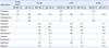
The resistance rates of clindamycin, erythromycin, chloramphenicol, and levofloxacin in 26 GBS isolates were 11.5%, 11.5%, 3.8%, and 7.7%, respectively. All isolates of GBS were susceptible to penicillin, ceftriaxone, and vancomycin. The cultures of M. hominis derived from 2 patients were resistant to erythromycin, tetracycline, ciprofloxacin, ofloxacin, clarithromycin, and azithromycin and susceptible to josamycin and pristinamycin. The M. hominis culture from 1 of the 2 patients was susceptible to doxycycline. The resistance rates to ciprofloxacin, ofloxacin, tetracycline, azithromycin, erythromycin, clarithromycin, and doxycycline for 56 U. urealyticum isolates were 96.4%, 55.4%, 19.6%, 12.5%, 8.9%, 7.1%, and 7.1%, respectively. All the U. urealyticum isolates were susceptible to josamycin and pristinamycin (Table 2).
DISCUSSION
The causal relationship between abnormalities in vaginal flora during pregnancy and PTB has gained a lot of attention [13-16]. Although this phenomenon is relatively common, the pathogenic mechanism that leads to S-PTB is still poorly understood. It is particularly difficult to define abnormal genital tract flora in pregnant women. The pathogenic role of specific microorganisms in the vagina as risk factors for S-PTB varies according to the investigators, because microorganisms commonly found in the lower genital tract are those that are most frequently isolated from patients with intrauterine infections. The colonization rate of microorganisms in the vagina could be affected by not only technical factors, such as detection method and sampling site, but also internal and external factors of each individual [17]. Possible racial/ethnic differences in the composition of the normal vaginal microflora also exist. Usui et al. [16] reported that absence of vaginal lactobacilli was a better predictor of S-PTB at <33 weeks of gestation than the presence of M. hominis, but its sensitivity and positive predictive value were no more than 28% and 25%, respectively. Breugelmans et al. [15] reported that there was no significant correlation between the presence of abnormal vaginal flora and S-PTB, and the risk of S-PTB increased when Ureaplasma species was associated with an abnormal vaginal flora.
Although BV has been considered a risk factor for S-PTB, investigators have been unable to prove a consistent association between BV and S-PTB [18]. In this study, clue cells were detected in only 1.4% of the S-PTB group, which suggests that BV is not associated with S-PTB. Possible explanations for our result are that BV is diagnosed mostly in the first trimester and the prevalence decreases in the second and third trimesters [18] and that the relationship of BV with S-PTB may vary according to the diagnostic criteria for the detection of BV.
U. urealyticum and M. hominis are among the organisms most frequently isolated from both placental membranes and amniotic fluid in patients with PPROM and those with PTL-WO [19-22]. Both organisms can initiate the synthesis of prostaglandins, resulting in S-PTB [9]. Nevertheless, there is much debate regarding whether genital mycoplasmas are pathogenic or simply frequent colonizers of the genital tract [14, 15, 20, 21]. The traditional view has been that genital mycoplasmas are part of the vaginal microflora in many women, and their presence in the lower genital tract, unlike their presence in the upper genital tract, has not been associated with an increased risk of S-PTB [22, 23].
Studies on the exact colonization rates of genital mycoplasmas should be performed to elucidate whether genital mycoplasmas are pathogenic. In our study population, overall detection rates of M. hominis and U. urealyticum in the vagina were 12.7% and 62.7%, respectively. The detection rate of M. hominis by PCR was higher than that by the culture method, while the detection rate of U. urealyticum by the culture method was higher than that by PCR. Possible explanations include lower sensitivity of the specific primer used in this study for the detection of U. urealyticum and/or false positive detection of Ureaplasma parvum by the culture method. A new species U. parvum (formerly biovar 1 of U. urealyticum serovars 1, 3, 6, and 14) was identified as U. urealyticum by using the Mycoplasma IST 2 test [24]. Lee et al. [14] reported that the prevalence of U. urealyticum was 28.6% (279/977) and that of M. hominis was 3.7% (36/977) in a vaginal fluid culture for genital mycoplasmas in pregnant women and that it was not associated with an increased risk for S-PTB. In contrast, Kafetzis et al. [25] reported that vaginal colonization with Ureaplasma species is associated with S-PTB because the rate of vertical transmission of Ureaplasma in mothers who experienced PTB was higher than that in mothers who experienced full-term delivery (33% vs. 17%). However, vaginal Ureaplasma colonization occurred among 36.5% of the mothers who experienced S-PTB and among 38% of the mothers who experienced full-term delivery. In this study, the RR of M. hominis and U. urealyticum in S-PTB to that in PTL-WO was 4.52 and 1.13, respectively, but these results were not statistically significant. Ureaplasma spp. can persist for weeks in the upper genital tract before spontaneous PTL or membrane rupture initiates S-PTB. Among the pregnant women who show Ureaplasma spp. and/or M. hominis colonization, only a small group will experience an ascending infection. The reason why this microorganism sometimes causes an ascending infection is not yet understood. There might be other associated factors that increase the capacity of the microorganism to invade the uterine cavity. These factors might include the host immune system, the virulence of the microorganisms, or local factors favoring the invasion of genital mycoplasmas.
GBS is detected in the vagina and rectum of 10-30% of pregnant women as normal flora, but it has emerged as a major pathogen for a variety of bacterial infections among pregnant women, non-pregnant adults, and elderly patients [26]. Garland et al. [27] reported that GBS is considered a risk factor for S-PTB because of its association with asymptomatic bacteriuria. However, when it colonizes the lower genital tract alone, it is not thought to promote preterm delivery. In this study, the RR of GBS in S-PTB was 2.26 (P<0.05), which suggests that GBS was not an independent risk factor for the development of S-PTB from PTL-WO.
N. gonorrhoeae and C. trachomatis are rarely implicated in uterine or fetal infection before the rupture of the membranes; however, both may have an adverse effect on pregnancy, thus increasing the risk for PTL [9]. N. gonorrhoeae has been shown to be present in approximately 1% of pregnant women and if left untreated, leads to a 2-5% RR of S-PTB [28]. In our study, the prevalence of C. trachomatis and N. gonorrhoeae by PCR was 2.4% and 0%, respectively; HSV I, M. genitalium, T. vaginalis, and T. pallidum were not detected using PCR.
Antibiotic administration to patients with PPROM is associated with prolongation of pregnancy and reduction in the rate of clinical chorioamnionitis and neonatal sepsis. The benefit has not been demonstrated in patients with PTL-WO and intact membranes. Major efforts are required to determine why some women develop an ascending intrauterine infection and others do not and which interventions reduce the deleterious effect of systemic fetal inflammation. According to a large randomized study, erythromycin treatment of pregnant women with U. urealyticum vaginal colonization does not confer any advantage in terms of reduction in the rate of S-PTB [29]. In this study, the resistance rate of erythromycin in U. urealyticum was 8.9%, while that of ciprofloxacin was 96.4%.
Our results demonstrated that genital mycoplasmas and GBS in the lower genital tract were not risk factors for the development of S-PTB from PTL-WO, although detection rates of those organisms in S-PTB were higher than those in PTL-WO. Differences in host response to the presence of genital mycoplasmas and GBS, rather than the mere presence of the bacteria themselves, may contribute to an increased risk of S-PTB.
The limitations of this study were that only a small number of PTL patients were enrolled and that the control group that did not experience PTL or PTB was only tested for GBS colonization. Additional large-scale studies on the colonization rates of GBS, M. hominis, and U. urealyticum in normal preterm women without labor or delivery are required to elucidate the pathogenic roles of these organisms in PTL-WO and S-PTB.
 XML Download