

 PDF
PDF ePub
ePub Citation
 Print
Print


Dear Editor,
Amyloidosis is a heterogeneous group of diseases characterized by extracellular amyloid deposits in different organs [1]. Deposition may occur secondary to a systemic disease or as primary amyloidosis in absence of a systemic disease. Periocular and orbital amyloidosis are rare, especially in the lacrimal apparatus. In South Korea, only 12 cases of amyloidosis have been reported, and these mainly concerned involvement of conjunctiva or cornea [2]. This is the first Korean report of a patient with systemic amyloidosis involving the lacrimal sac.
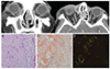
An 81-year-old man was referred to our hospital with complaints of tearing in both eyes, tenderness, and swelling in the region overlying the right lacrimal sac. He had a hemato-oncological history of diffuse large B cell lymphoma of the left kidney and a recent history of amyloidosis of the stomach but no ophthalmic disease history. On physical examination, best-corrected visual acuity was 0.5 in the right eye and 0.4 in the left. There was mucoid reflux upon compression, and a soft mass was detected by palpation in the right lacrimal sac area. The tear meniscus was elevated, and impaired clearance of fluorescein dye was noted from the tear film in both eyes. The remainder of the ophthalmic examination was normal. Facial computed tomography revealed swelling of the right medial canthal area and dilatation of both lacrimal sacs with peripheral enhancement of fuzzy margins (Fig. 1A). The patient underwent endoscopic dacryocystorhinostomy in both lacrimal sacs. During the operation, a cheese-like yellowish mucoid substance was noted on the left lacrimal sac. A biopsy of the left lacrimal sac was performed, and we removed all of the yellowish material. Some days afterward, histological examination of the biopsy sample showed amorphous and eosinophilic substances by hematoxylin & eosin staining and orange-colored amyloid deposits by Congo red staining, the latter of which exhibited apple-green birefringence under a polarized light microscope (Fig. 1B-1D). All of these histological findings corresponded to amyloidosis; thus, a diagnosis of amyloidosis was made.
The various forms of amyloidosis are described as localized or systemic, primary or secondary, or heredofamilial. Amyloid proteins can be derived from many sources, including immunoglobulin lambda or kappa light chains, protein AA, protein AP, some proteins of prealbumin origin, or transthyretin [3]. With respect to immunoperoxidase staining, two subunits are pertinent to the subject of orbital amyloidosis. Protein amyloid AA, derived from serum protein A, is associated with amyloidosis previously considered to have arisen secondary to chronic inflammatory or infectious disorders, such as tuberculosis, leprosy, rheumatoid arthritis, or osteomyelitis. The other protein, amyloid AL, consists of all or part of the immunoglobulin lambda or kappa light chain and is common to cases of primary amyloidosis, which usually occurs secondary to a benign, low-grade, light chain-producing monoclonal gammopathy and is associated with widespread organ deposition and dysfunction [45]. Serum electrophoresis in our patient did not show monoclonal gammopathy but rather an increased gamma fraction; thus, given the lack of a systemic chronic inflammatory and infectious disorder, primary systemic amyloidosis was diagnosed. Unfortunately, no further analysis was performed to define the amyloid type, which is a limitation of this case report.
The modalities used to treat primary localized amyloidosis depend on disease location; for example, surgical debulking or combined surgical debulking with external beam radiation is used to treat orbital amyloidosis, liquid nitrogen cryotherapy to treat conjunctival amyloidosis, and some cases receive only observation [1]. Isolated orbital and adnexal amyloidosis are usually treated by surgical removal of the amyloid mass. However, local recurrence is not uncommon in such cases [4]. In our case, we did not microscopically confirm eradication of the amyloid substance during the operation. Thus, close observation is ongoing for amyloidosis recurrence. Surgical excision should be planned if the disease relapses.
In summary, we report a case of lacrimal sac amyloidosis. To our knowledge, this is the first report on primary amyloidosis involving the lacrimal sac.
 XML Download