

 PDF
PDF ePub
ePub Citation
 Print
Print


Dear Editor,
Vogt-Koyanagi-Harada (VKH) disease usually presents as bilateral panuveitis associated with poliosis, vitiligo, alopecia, and central nervous system and auditory signs. Simultaneous onset in both eyes is common. In unilateral cases, the second eye is typically affected within 2 weeks of the first eye. We describe the case of a patient with unilateral VKH in which relapse was accompanied by posterior scleritis after tapering systemic steroid treatment.
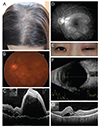
A 37-year-old woman visited our clinic with blurred vision in her left eye. She had suffered from severe headache with tinnitus for 1 week, left eye pain for 4 days and metamorphopsia for 1 day. She also complained of a several month history of alopecia (Fig. 1A). The initial best-corrected visual acuity (BCVA) was 1.0 in the right eye and 0.16 in the left eye. The initial intraocular pressure was 10 mmHg in both eyes. A mild anterior chamber reaction was observed without keratic precipitate. On fundus examination of the left eye, serous retinal detachment was observed, but no vitreous cells or haziness were detected (Fig. 1B-1D). The patient was diagnosed with unilateral VKH disease, and prescribed prednisolone 60 mg/day. She was also administered topical prednisolone and cyclopentolate eye drops. Four weeks later, all of her symptoms had resolved completely and the BCVA of the left eye had recovered to 1.0. Thus, we began to taper systemic steroid from 60 to 5 mg over 2 months.
After 3 weeks, periorbital swelling, chemosis, conjunctival injection and itching sensation occurred in the left eye, appearing as acute allergic conjunctivitis (Fig. 1E). Neither periorbital pain nor serous retinal detachment was reported. One day later, symptoms were aggravated; although BCVA was still 1.0 in the left eye, serous retinal detachment had developed, though to a less severe degree than on initial presentation. Ultrasonography of the left eye revealed increased thickness of the posterior scleral wall and typical T sign (Fig. 1F and 1G), which is caused by the spread of inflammation along Tenon's space into the optic nerve sheath. We recommended that she be hospitalized and start high-dose intravenous steroid pulse therapy (methylprednisolone 1 g/day for 3 days). After starting high-dose steroid pulse therapy, the patient's general condition and ocular/visual symptoms began to improve. Now she stopped steroid therapy and in the past several months, there have been no other complications or sequelae.
VKH is a non-necrotizing diffuse granulomatous inflammation involving the uvea. Although the exact cause of inflammation remains unclear, current evidence suggests that it involves an autoimmune process driven by T lymphocytes associated with melanocytes. The differential diagnosis of VKH disease includes sympathetic ophthalmia, uveal effusion syndrome, posterior scleritis, acute posterior multifocal placoid pigment epitheliopathy, and sarcoidosis. VKH disease usually presents as bilateral panuveitis. Simultaneous onset in both eyes is common, and, in unilateral cases, the second eye is typically affected within 2 weeks of the first.
There have been few reports of unilateral VKH disease. A et al. [1] reported the case of a 4-year-old boy with unilateral VKH disease; even though there was no inflammation in the unaffected eye in that case, ultrasonography revealed thickening of the choroids in both eyes.
Posterior scleritis is a relatively rare disease that occasionally appears with exudative retinal detachment and multiple pinpoint leaks on fluorescein angiography, similar to the signs of VKH disease. Furthermore, posterior scleritis can be differentiated from orbital cellulitis based on complaints of lid swelling, erythema and eye pain in scleritis. Orbital cellulitis and posterior scleritis are both potentially life-threatening conditions that require urgent management [23].
Differentiation between VKH disease and posterior scleritis is achieved through observation of sun set glow fundus and neurologic or dermatologic signs not seen in posterior scleritis. Posterior scleritis is also usually unilateral, and T-sign is a unique characteristic of this disease. Recently, there have been some reports, especially in Japan, of posterior scleritis appearing concurrently with VKH disease in patients; Kouda et al. [4] reported that posterior scleritis was an early manifestation of VKH.
In our case, neurologic symptoms such as headache and tinnitus appeared as clinical features of VKH, while T-sign and lid swelling were signs of posterior scleritis. Anterior chamber reaction and serous retinal detachment were also observed in both VKH and posterior scleritis. Thus, we conclude that our case involved unilateral VKH disease with posterior scleritis.
 XML Download