

 PDF
PDF ePub
ePub Citation
 Print
Print


The SCORE (Standard Care vs Corticosteroid for Retinal Vein Occlusion) study revealed the benefit of steroid treatment in central retinal vein occlusion (CRVO) associated with macular edema (ME) [1]. Herein, we report a case of the reversal of early CRVO with optic disc swelling after intravitreal dexamethasone implant (Ozurdex; Allergan Inc., Irvine, CA, USA) injection. This case suggests that early intravitreal dexamethasone implantation may be a good treatment option in CRVO with co-existing optic nerve swelling. This treatment could relieve optic nerve edema causing the occlusion of venous flow, thereby improving venous outflow.
Case Report
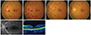
A 44-year-old female without any previous medical history visited our clinic because of the sudden onset of intermittent blurred vision in her left eye. Her best-corrected visual acuity (BCVA) was 20 / 22 and fundus examination revealed multiple retinal, preretinal, and subretinal hemorrhages throughout the retina with tortuous retinal veins in her left eye (Fig. 1A). The right eye showed a completely normal fundus. Fluorescein angiography revealed delayed retinal venous filling. Arteriovenous transit time was slightly prolonged to 19 seconds and there was faint leakage in the late phase, but no signs of any vascular non-perfusion (Fig. 1B). Cardiologic and neurologic work-up and laboratory tests (homocysteine, protein C and S, fibrinogen, anticardiolipin antibodies, and lupus anticoagulants) were normal except for weakly positive anticardiolipin IgM and mild iron deficiency anemia. The patient denied any valsalva episodes. We decided to observe the patient for the following two weeks without making a diagnosis. Two weeks later, her BCVA was slightly decreased to 20 / 25, and an increased number of retinal hemorrhages with severe disc swelling were noted (Fig. 1C), but there was still no sign of ME (Fig. 1D). At this time, the patient preferred to undergo medical intervention to improve her subjective symptom. We discussed with her that the drainage site of the retinal vein is located in the lamina cribrosa within the optic nerve and an intravitreal dexamethasone implant may help alleviate the progression of the vein occlusion by reducing optic nerve edema. The patient agreed to the treatment and an intravitreal dexamethasone implant 0.7 mg was injected.
Five days later, her BCVA was 20 / 22 and there were improvements in disc swelling and retinal hemorrhage (Fig. 1E). One month later, her BCVA improved to 20 / 20 and her subjective visual symptom was completely improved. Fundus examination revealed marked improvement of retinal hemorrhages and vascular tortuosity along with almost complete resolution of disc swelling (Fig. 1F).
Discussion
A compartment syndrome at the site of the lamina cribrosa is thought to be the key mechanism in the pathophysiology of CRVO. The current treatment strategy for CRVO is typically directed to complications such as ME or neovascularization rather than the venous occlusion itself. Although many studies have already shown the benefits of anti-vascular endothelial growth factors or steroids for the treatment of CRVO-associated complications [2,3], this case suggests that even in early CRVO, if associated with optic nerve swelling, steroid treatment could decrease disc swelling, thereby relieving compartment obstruction and improving vascular hemorrhage. Steroids may reduce the disc swelling, which subsequently decreases the pressure of the scleral ring and may improve the venous outflow. This may potentially change the natural course of disease progression of CRVO and its various subsequent complications.
 XML Download