

 PDF
PDF ePub
ePub Citation
 Print
Print


Superior oblique palsy (SOP) is the most common isolated cranial nerve palsy induced by trauma, idiopathic, vascular insufficiency, tumor, or iatrogenic factors [1]. The superior oblique muscle has a unique anatomical function because the anterior fibers impart a primarily excyclotorsional force on the globe, while the posterior fibers impart primarily depressing and abducting forces on the globe. For these reasons, patients with unilateral superior oblique muscle palsy generally evidence hypertropia and excyclotorsion of the paretic eye.
SOP is diagnosed by clinical observations of ocular versions, measurements of misalignment of ocular position at the nine cardinal gazes, and head tilt positions [1,2]. Moreover, cyclotorsion is a crucially important feature in SOP. The double Maddox rod test (DMRT), fundus photography, major amblyoscope, and Lancaster test are all regarded as useful tools to evaluate cyclotorsion. These methods may reveal the torsional angle in a subjective or objective manner. The discrepancies among measurements of cyclodeviation have been well established. Na et al. [3] reported that both excyclotorsion and hypertropia showed a significant and positive correlation with inferior oblique overaction (IOOA). Hypertropia and excyclotorsion showed a weaker positive correlation in congenital monocular SOP. However, Na et al. [3]'s report included patient populations with congenital and acquired palsies.
Therefore, we aimed to evaluate the characteristics and the correlation between hypertropia and excyclotorsion in acquired unilateral superior oblique muscle palsy via subjective measuring tests including the DMRT and major amblyoscope, and an objective measuring test involving fundus photography.
Materials and Methods
Clinical charts of 31 patients diagnosed with acquired unilateral superior oblique muscle palsy during March 2007 and February 2009 were retrospectively reviewed. This research study was reviewed and approved by the institutional review board of Kim's Eye Hospital. The study included patients with acquired unilateral SOP who were diagnosed by a three-step test. Exclusion criterion were as follows: 1) previous extraocular surgery history; 2) any suspected sign of other paralytic strabismus except SOP; 3) presence of bilateral SOP which was defined by right eye hypertropia in left gaze; 4) left hypertropia in right gaze; and 5) positive Bielschowsky head tilt test to either side.
Every patient had a full ophthalmic examination including a visual acuity test and slit-lamp examination. Patients were divided into two groups (concordance group and discordance group) according to the correspondence between the hypertropic eye (paralytic eye) and the more extorted eye (non-fixating eye), as evaluated via fundus photography. For example, if a patient who had a right hypertropia showed a marked excyclotorsion in the right eye, the patient was classified into the concordance group. All data were collected at the first visit.
For the evaluation of hypertropia, an alternate prism cover test was conducted using a 5 m far target in the primary position, up and down gaze, lateral gaze, and bilateral head tilt positions of 30°. IOOA was quantified via the duction version test from grades 1 to 4. Inferior oblique overaction was graded as 1 if the adducted eye was 1 mm above the inferior margin of the fellow eye pupil, grade 2 if it was 2 mm above, grade 3 if it was 3 mm above, and grade 4 if it was 4 mm above.
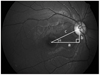
We employed two subjective methods (DMRT and major amblyoscope) and one objective method (fundus photography) to quantify the cyclotorsion. The DMRT was conducted as followed. Red Maddox rods were placed before both eyes, and a Maddox rod was turned until the two lines were perceived as parallel. In this method the angle of rotation determines subjective excyclotorsion. Another method for measuring subjective excyclotorsion uses the major amblyoscope (ED-011; Takata Ophthalmic Instruments, Tokyo, Japan). Two slides (ST1 and ST2) were used. One slide featuring a vertical line was placed before the non-paretic eye, and another slide marked with a horizontal line was placed before the paretic eye. Patients were asked to describe the angle of the two lines. The investigator rotated the arms of the major amblyoscope until no cyclodeviation was perceived. The required degree of rotation of the slide by the investigator was used as the measurement of cyclodeviation. Fundus photography was conducted using a digital fundus camera (Canon CR6-45NM with Canon EOS 20D; Canon, Tokyo, Japan) with internal fixation. A horizontal line was drawn from the center of the optic disc to the fovea, and the other line was drawn horizontally from the fovea. Usually, the fovea is located slightly inferior in relation to the optic nerve. The angle made by the two lines was the disc foveal angle, and that angle was expressed in terms of tangential value (tan α = b/a) (Fig. 1). This value represented objective excyclotorsion in each eye. Ocular torsion measured by fundus photography was evaluated by the total excyclotorsion, which was the difference between the two eyes. For example, if fundus photography showed a 15° excyclotorsion in the right eye and an 8° excyclotorsion in the left eye, the total excyclotorsion is defined as 7° (15° to 8°).
Data were analyzed using SPSS ver. 14.0 (SPSS Inc., Chicago, IL, USA). The results from quantitative variables were expressed as the means ± standard deviation, and comparisons between groups were conducted using non-parametric Mann-Whitney and Friedman tests. Correlations between tests were assessed via non-parametric Kendall's tau rank correlation. A p-value of <0.05 was regarded as statistically significant.
Results
Data from 31 patients were included in this study. The median age at presentation was 52 years (range, 9 to 75 years) and three of the 31 patients were female.
Vertical and horizontal deviation
The mean degree of hypertropia was measured as 5.83 prism diopters (PD) in primary gaze. Horizontal deviation was observed in 12 patients (38.7%). Among these 12 patients, ten showed an exodeviation (mean value, 9.3 PD; exodeviation more than 8 PD, five patients) and two patients exhibited an esodeviation (6 PD and 9 PD, respectively). Upon presentation, 20 (64.51%) patients evidenced inferior oblique overaction, with a mean grade of 1.45.
Relationship between objective torsion and subjective torsion
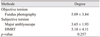
The mean value of objective torsion was 5.09° ± 3.84°. The subjective excyclotorsion degrees were 5.18° ± 4.11° suggested by the DMRT and 3.65° ± 1.93° using the major amblyoscope. We detected no significant differences among tests (Friedman's test, p = 0.257) (Table 1). The degree of torsion measured via fundus photography and the amplitude of the major amblyoscope measurements had a positive relationship. However, this was not a significant relationship (correlation coefficient = 0.216, p = 0.293). Although the degree of torsion in fundus photography and the degree of torsion measured by the DMRT had a negative relationship, these results were not significant (correlation coefficient = -0.122, p = 0.422).
Relationship between hypertropia and excyclotorsion
The degree of hypertropia and the degree of torsion measured using fundus photography were not significantly correlated (correlation coefficient = 0.249, p = 0.072). The degree of hypertropia and the amplitude of subjective excyclotorsion, as measured by the major amblyoscope and DMRT, also evidenced no significant relationship (correlation coefficient = 0.108, p = 0.591 and correlation coefficient = -0.124, p = 0.424).
Concordance group and discordance group
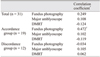
Nineteen of 31 patients comprised the concordance group (61%). No significant differences were noted between the concordance and the discordance groups (Table 2). In the concordance group, a significant positive correlation between hypertropia and excyclotorsion as determined via fundus photography was detected (p = 0.011). However, there was no significant correlation between hypertropia and torsional angles in the discordance group, according to measurements conducted via the three different methods (Table 3).
Discussion
There are various methods by which the degree of excyclotorsion can be determined [4]. The relevant methods were categorized into objective and subjective methods. We employed fundus photography for objective torsional measurements and major amblyoscope and DMRT as subjective measurements. The objective and subjective methods were not correlated. With regard to the subjective methods, the major amblyoscope results were positively correlated with the objective results, as compared with the DMRT, which evidenced a negative correlation. However, none of these results were significant. Seventy-three percent of patients evidenced excyclotorsion in the DMRT, and the mean degree of excyclotorsion was 5.18°. The excyclotorsion measured by the amblyoscope was 3.65°. These results were similar to those of a previous study by von Noorden et al. [5] who reported that excyclotorsion measured via the DMRT occurred in 76% of patients with unilateral SOP, with a mean degree of excyclotropia of 3.5° [5].
The cyclotorsion of fundus photography was similar to that seen in the DMRT results, and was larger than the subjective torsion (fundus photography 5.09°, DMRT 5.3° and major amblyoscope 3.9°). These findings demonstrate the similarity to the results reported by Ruttum and von Noorden [6]. They suggested that discrepancy in the testing results may have been due to fusional function. It may also result from diverse fusional power in various situations. Fusional power may contribute to the diminution of the torsional angle and result in the reduction of torsional deviation in the major amblyoscope. Therefore, the angle measured using the major amblyoscope was the smallest among the tests.
Hypertropia is induced by inferior oblique overaction of the paretic eye. Inferior oblique overaction also causes excyclotorsion, and alternatively, pseudo-overaction of SOP of the nonparetic eye can cause hypertropia [7-9]. In the relationship between hypertropia and excyclotorsion, 19 patients showed hypertropia as a demonstration of this relationship. Olivier and von Noorden [10] reported that approximately 75% of these cases were attributable to a coincidence due to fixation preference. Our results were slightly lower than in a previous study. This may be because all the cases in the present study were an acquired form. Patients with acquired SOP may more commonly have a fixation preference than patients with congenital SOP, and thus the patients may continue to have a fixation preference after developing SOP. In this study, the relationship between hypertropia and excyclotorsion evidenced no significant correlation among patients. However, objective excyclotorsion, which was evaluated via fundus photography, showed a significant positive correlation in the concordance group after patients were divided into two groups according to fixation preference. If patients in the discordance group fixated with their paralytic eye, a significant correlation would be noted between objective excyclotorsion and hypertropia. As a consequence, in the case of acquired SOP with patients who fixate using their nonparalytic eye, the objective excyclotorsion method may be superior to subjective methods. Because the angle of deviation is largely measured according to Herring's law in the discordance group, patients who fixate with the paralytic eye may not have accurate measurements.
In terms of finding no correlation between hypertropia and excyclotorsion, anatomical factors may be relevant to the characteristics of SOP. Mollan et al. [11] recently identified 133 cases of SOP. Among the unilateral isolated cases, 38.3% were considered congenital, 29.3% followed trauma, and 23.3% were presumed to be vasculopathic in origin, while no cause could be established in 7.5%. Based on their results, more than half of the cases of acquired unilateral SOP were attributable to trauma or vasculopathy. We suggested that the selective attack of the superior oblique muscle fiber may result in dominant hypertropia or dominant excyclotorsion. If the mechanical damage or vascular ischemia involves the anterior fiber, excyclotorsion may be the result. Conversely, if the damage or ischemia affects the posterior fiber, hypertropia may develop.
In conclusion, the relationship between the amount of excyclotorsion and hypertropia in acquired SOP is weak. But in the concordance group, a significant positive correlation between hypertropia and excyclotorsion was expressed. Knowing the relationship of objective torsion and subjective torsion in patients with acquired SOP, and fixation preference can help understand acquired SOP and help predict the surgical results and operative quantitative measurements.

 XML Download