

 PDF
PDF ePub
ePub Citation
 Print
Print


According to the definition by the World Health Organization (WHO), a person with low vision has impairment of visual functioning even after treatment and/or standard refractive correction, and has a visual acuity of less than 6 / 18 to light perception or a visual field less than 10 degrees from the point of fixation, but who uses, or is potentially able to use vision for the planning and/or execution of a task for which vision is essential [1]. Rehabilitation of low vision is becoming more popular as low vision patients strive to complete daily tasks or to work by utilizing their residual vision and remaining sight. The total number of registered people with low vision in Korea was reported as 216,881 by the Minister of Health and Welfare in 2007. However, the actual number may be higher as current estimates suggest low vision patients comprise 0.8% of the population in developed countries. Consequently, rehabilitation for patients with low vision is becoming increasingly important.
Low vision rehabilitation can be categorized in two ways. One method utilizes low vision aids while the other method focuses on the training of visual function to maximize a patient's residual vision. According to a study of 500 low vision patients in Korea, more than half of low vision patients are aware of low vision aids; however, they are unaware of the possibility of visual function training [2]. Low vision rehabilitation using low vision aids is very useful in increasing near and distance visual acuity and improving the quality of life in low vision patients [2-6]; however, expense is a major obstacle to the widespread use of low vision aids. Most low vision aids are quite costly and the potential tax benefits are not substantial enough to make the purchase economically feasible for many low vision patients. Additionally, many low vision patients in Korea have lower incomes. Finally, low vision rehabilitation with visual function training is essential for the efficient use of low vision aids.
Eccentric viewing (EV), a representative method of visual function training, consists of using any non-foveal point on the retina for viewing [7]. The fovea is located at the center of the macula and is the core area used for fixation in the healthy retina; however, when the central retina is damaged, the eye must utilize a new retinal area for viewing. A previous study reported that during EV training [8], a preferred retinal locus (PRL) can develop to replace the fovea and has since been suggested to be the most important factor in EV training [9-11]. Research on monkeys with bilateral laser-induced foveal lesions showed they developed a new PRL within days, although weeks to months were required before they were able to train their eye movements to accurately place targets on their PRL [12]. However, in practice many patients who already view eccentrically do not realize they are not fixating on the target. In fact, many patients, particularly older patients or those with sudden onset visual field restriction do not naturally adapt a PRL and need to be educated that they have a scotoma and can view around it.
Low vision specialists have studied various methods and techniques for training patients who have not adapted to their central scotomas to view eccentrically; however, how to best optimize EV training techniques is still not fully understood. In addition, EV training is relatively unfamiliar compared to the application of low vision aids, especially in Korea. However, as the standard of living in Korea develops, the desire for better vision is increasing. Additionally, as the population ages, the number of patients with central scotomas is larger because of diseases such as age-related macular degeneration (AMD). Consequently, EV training could be a useful technique for low vision rehabilitation. To investigate the clinical effects of EV training for low vision rehabilitation in patients with central scotomas, we analyzed the association between PRL and direction of EV, changes following EV training, and factors related to the success of EV training. Our findings may help to establish the feasibility of EV as a useful technique for low vision rehabilitation.
Materials and Methods
Thirty low vision patients (16 females, 14 males) with central scotomas were recruited from a local clinic. All patients had visual field defects within the central 10 degrees in the dominant eye. However, patients with multiple remaining islands of their visual field or prior experiences with low vision rehabilitation were excluded to avoid misleading results, even if they had central scotomas within the central 10 degrees. Patients with significant media opacities and/or other ocular diseases that could affect visual function were also excluded. The Institutional Review Board approved the protocol and consent form for this study (IRB no. C2010014 [309]). All procedures were performed under the tenets of the Helsinki Declaration and all patients gave their informed consent.
Different examiners were assigned to various tests to prevent information bias and each test was performed by only one examiner to maintain consistency. The technique used to determine the direction of EV involved the patient sitting in order for an optotype with a 20 / 225 size of a Feinbloom distance chart to be at eye level and 1 m away. The patient was then instructed to slowly look up, down, left, and right and comment on which direction yielded the clearest image. The patient was then instructed to move their viewing direction circumferentially two clock hours at a time, comparing the new direction to the previous best of all tested viewing directions. Once a single direction yielding the best possible vision was identified, the patient was instructed to practice using that visual direction.
The location of a patient's PRL was evaluated with a direct ophthalmoscope (Welch Allyn Inc., Skaneateles, NY, USA) by having the patient identify and fixate on a target transmitted through the fixation aperture of the direct ophthalmoscope. A 50° fundus photograph was obtained with a fundus camera (TRC 50IX; Topcon, Tokyo, Japan) by having the patient fixate on a flickering point. The Humphrey Field Analyzer II (Carl Zeiss Meditec, Dublin, CA, USA) and Goldmann kinetic perimetry (Topcon) were used to study the location, sensitivity, and fixation stability of the PRL. For both field tests, a white V 4e test light (stimulus size 64 mm2, intensity 10 dB) moved at a speed of 4°/sec. A patient's visual fields were tested in 12 meridians evenly spaced 30° apart (0°, 30°, 60°, 90°, 120°, 150°, 180°, 210°, 240°, 270°, 300°, and 330°). In screening tests, the Humphrey Field Analyzer is used primarily for its automation and standardization [13]. However, the movement direction of the test light (periphery to center) was not appropriate for evaluating the central scotomas in the present study; therefore, the Humphrey field analyzer was used in combination with Goldmann kinetic perimetry in which the test light moves in the opposite direction (central to periphery).
The direction of EV, the location of the PRL, and the preserved visual field were divided into 8 categories (Table 1) and the results of each test were marked in the appropriate category. Accordance between all tests of EV direction and PRL was regarded as a coincidence. Once the best direction of EV and the location of the PRL were identified, the patient and their guardians were educated and trained to intentionally use EV while looking at a target at 40 cm and 1 m for at least 15 minutes a day at each distance. In addition to the 30 minutes of daily training, patients were encouraged to use EV in their daily life.
After 2 weeks, the proportion of patients who continued to practice using EV was assessed. The effect of EV training was evaluated in the continuance group using best-corrected visual acuity (BCVA), reading speed, and a satisfaction questionnaire. For standardization, BCVA for distance was checked at 3 m distance from a Feinbloom distance chart with a luminance of 500-600 lx and at 40 cm from a Precision vision near chart with a luminance of 700-800 lx for near vision. The results were converted to the logarithm of the minimum angle of resolution (logMAR) scale. Reading speed was evaluated under the same conditions used for near vision testing utilizing sentences designed by the low vision clinic. The sentences were composed of a 20 point size font using the Batang style of the Korean alphabet. Reading speed was recorded in letters per minute and misread words were not counted. The satisfaction questionnaire contained 10 items selected from the Korean version of low vision quality-of-life questionnaire [14,15]; each item was graded on a 6-point scale. Patients were instructed to grade the items between 5 (no difficulty with the item because of vision) and 1 (great difficulty with the item because of vision) or as 0 (item no longer performed because of vision). The range of total scores from 0 to 50 (a higher score represented a greater degree of satisfaction) was used to assess the patient's level of satisfaction.
Data were recorded and analyzed using SPSS ver. 16.0 (SPSS Inc., Chicago, IL, USA). Comparisons between the accordance and discordance groups and between the continuance and discontinuance groups were assessed using the Mann-Whitney U-test. The Wilcoxon signed-rank test was used to compare changes in visual function in the continuance group following EV training. The probability level for statistical significance was set at 5%.
Results
Of the 30 subjects, 18 (60%) had a diagnosis of atrophic AMD, 4 had end-stage glaucoma, 4 were post-trauma, 2 had macular dystrophy, and 2 had retinopathy of prematurity (ROP). The mean age was 50.3 ± 17.7 years with a range of 16 to 86 years. The mean far BCVA, near BCVA, reading speed, and questionnaire score were 1.01 ± 0.37 (logMAR), 0.8 ± 0.34 (logMAR), 26.4 ± 7.7 (letters/min), and 25.1 ± 11.4 (score), respectively.
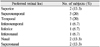
Half of the patients (15) were in accordance regarding their direction in EV, PRL, and visual field (accordance group). The majority of PRLs were located in the temporal and superotemporal retina (Table 2). There was no difference in distribution of disease (AMD, 9; trauma, 2; glaucoma, 2; ROP, 1; and macular dystrophy) between the accordance group and the discordance group (Table 3) and no significant differences in demographics and basic visual characteristics such as mean age, far BCVA, near BCVA, reading speed, and satisfaction questionnaire score between the two groups (Mann-Whitney U-test, p > 0.05). The ratio of men to women was higher in the accordance group (8:7) than in the discordance group (6:9). Although the proportion of continuous EV was higher in the accordance group (73.3%), more than half (60%) of the subjects in the discordance group used EV continuously (Table 4).
Two-thirds of the patients (20) reported using EV continuously after 2 weeks of training (continuance group). The disease distribution in the group included 13 patients with AMD, 3 with a history of trauma, 1 with glaucoma, 1 with macular dystrophy, and 2 with ROP. The disease distribution in the discontinuance group (10) included 5 patients with AMD, 1 with a history of trauma, 3 with glaucoma, and 1 with macular dystrophy (Table 3). There were no significant differences in demographics and basic visual characteristics such as mean age, ratio of men to women, far BCVA, near BCVA, reading speed, and questionnaire score between the continuance and discontinuance groups (Mann-Whitney U-test, p > 0.05). In the continuance group, more than half (55%) of the subjects were in accordance with the EV direction, PRL, and visual field; in contrast, less than half (40%) of the subjects in the discontinuance group were in accordance with the same parameters (Table 5).
Following 2 weeks of EV training, far and near BCVA did not significantly improve (0.1 logMAR on average); however, mean reading speed and questionnaire score did significantly increase after EV training (Wilcoxon signed-rank test, p < 0.05). The mean reading speed improved from 26.0 ± 8.0 to 54.0 ± 11.0 letters per minute (p < 0.001) and the mean questionnaire score increased from 25.0 ± 10.0 to 30.0 ± 12.0 (p = 0.025) (Table 6).
Discussion
The present study identified a convenient and inexpensive method for detecting the direction of EV and quantified clinical improvements in functional vision and patient satisfaction following EV training. EV training can be used as an effective method for low vision rehabilitation of central scotomas instead of the existing optical aids-based Korean low vision rehabilitation standard. Macular diseases such as AMD, cone-dystrophy, macular myopic degeneration, vitelliform dystrophy, post-traumatic macular scarring, and Stargardt are characterized by the development of a central scotoma. Besides reducing reading speed, a central scotoma interferes with other visual functions including space perception, contrast sensitivity, stereopsis, and fixation stability [9,10]. The WHO estimates that 8 million people worldwide are severely visually impaired because of AMD [1]; furthermore, according to a study of low vision patients in Korea [2], AMD is the most frequent cause of low vision in adults older than 20 years. As the prevalence of macular diseases continues to increase, a growing concern regarding central scotomas and the PRLs of the patients has led to increased investigation regarding the improvement of visual performance; EV is a representative method of visual function training.
Generally, central visual field could be preserved until the terminal stage of glaucoma in most patients. However, in the present study, in 4 glaucoma patients, the preserved visual field was the temporal island rather than central field which was burned out. Two patients with a diagnosis of ROP in the present study had central scotomas within the central 10° on the visual field test, and the obvious cause of the central scotomas could not be determined. Therefore, the disease categories of glaucoma and ROP were included in the present study, although not sufficiently apparent to be categorized as macular disease.
Eccentric PRLs have been known to exist for many years and develop in approximately 84% of eyes affected by central scotomas [16]. However, the factors determining the development of a PRL at a precise location relative to the scotoma and their characteristics are still not well understood. Previous studies have reported that PRLs were more frequently located relative to a specific location of the scotoma, although, the reports regarding the precise location are conflicting [16-18]. Additionally, studies have shown the location of PRLs can change as a function of the type of macular disease present, the size of the target, the luminance level of the background, and the functional task [19-24].
In the present study, PRLs were preferentially located in the temporal (20%) and superotemporal (20%) retina in relation to the fovea in the accordance group; however, the locations of the PRLs in the accordance group were highly variable (Table 2) and a closer analysis of PRLs in the discordance group was limited. Three explanations are proposed for the discrepancy as there were no significant differences between the accordance and discordance groups with respect to demographics and basic visual characteristics.
First, the discrepancy can be explained by the limitations of the technique used to detect the direction of EV and the location of the PRL. Often PRLs are located adjacent to the atrophic area causing the central scotoma [16,25]. Recent studies have used various types of scanning laser ophthalmoscopes (SLOs) and microperimeters that offer new possibilities for evaluating EV in a more precise manner. The evaluation is performed using simultaneous video magnification of the retina which allows for rapid recording of the fixation stability with digital fundus imaging and a visual field sensitivity measurement [26-29]. However, in the present study, the direction of EV and the location of the anatomical PRL could be inaccurate as they were evaluated by passively moving the fixation target. Additionally, the visual stimulus of the direct ophthalmoscope could not be standardized. Further evaluation using a more precise method such as an SLO and microperimeter, may help clarify the relationship between the direction of EV, the location of the PRL, and the preserved visual field.
Second, patients with central scotomas may use more than 1 PRL and their location can vary depending on the viewing conditions. Multiple remaining islands of visual field were excluded in the present study; however, the possibility of multiple micro PRLs that could not be detected by a conventional visual field analyzer still exists. The patients may use a tiny area in an indentation in the retinal lesion to identify a small letter with reasonable acuity, but a larger and more peripheral area when looking at larger objects. Additionally, several patients have reportedly used 2 different retinal loci for different lighting conditions [18,21,24,30,31]. Furthermore, Deruaz et al. [32] reported on the adaptation of more than 1 PRL through a training procedure that developed a newly selected trained retinal locus in addition to the initial spontaneously developed PRL. In the present study, the existence of multiple PRLs could have caused the discrepancy between the direction of EV, the location of the PRL, and the preserved visual field.
Third, in some patients with a macular lesion, foveal function may be preserved. Using functional brain magnetic resonance imaging in macular degeneration, Baker et al. [33] documented large-scale reorganization of visual cortex processing occurs only in the complete absence of functional foveal vision. The researchers found no evidence for reorganization despite the clinical presence of PRLs in 2 individuals with extensive retinal lesions with some foveal sparing. Preservation and utilization, as long as functional foveal vision persists, can be a contraindication for EV training. In addition, the presence of foveal vision makes determination of the direction of EV and the location of a PRL difficult. However, as the macular lesion progresses, the complete loss of functional foveal vision can result and lead to an indication for EV training. The authors of the present study suggest careful evaluation and close follow-up to monitor the onset, status, progression, and characteristics of macular lesions.
EV training in low vision patients with central scotomas can significantly improve the efficiency of functional vision and the level of patient satisfaction. After EV training, reading speed doubled and the satisfaction questionnaire score increased by 9% in the continuance group; however, there was little to no improvement in far and near BCVA. Reading speed is a better parameter than visual acuity when reporting results of visual rehabilitation because reading is significantly more demanding than identifying a few optotypes on a visual acuity chart [27,32,34]. Importantly, the methods in the present study used to train and identify the direction of EV were inexpensive, in contrast to SLOs or microperimeters which are too expensive to allow practical application in low vision rehabilitation.
One-third of the patients in the present study did not use EV continuously; however, the clinical direction of EV was detected in all of the patients. There were no significant differences in demographics, causes of disease, or basic visual characteristics between the continuance group and discontinuance group or between the accordance group and the discordance group. Given the continuous sensitivity of maintaining EV (11 / 15, 73%) in the accordance group, a correlation between accordance and continuance of EV is suspected. However, the discontinuous specificity of maintaining EV was 40% (6 / 15) in the discordance group, indicating EV training was effective in 60% of patients in the discordance group. This result indicates agreement is not essential for effective EV training.
Maintenance of EV may be significantly associated with training techniques and conditions. Including SLOs and the microperimeter, many training techniques for EV have been introduced, such as prismatic scanning or relocation, strips and rotators with letters or numbers of various sizes, and specialized training sheets [35-37]. However, in the present study, EV training was entirely self-directed due to practical conditions that could have decreased the effect of EV training. For effective EV training, well-organized visual rehabilitation programs and environmental support are essential to support patients' efforts. The authors of the present study suggest experts in low vision rehabilitation take an active interest in EV and recognize low vision rehabilitation may differ significantly depending on both the characteristics of the individual and the disease.
Recent research on PRLs and reorganization of visual processing using functional magnetic resonance imaging shows EV may be related to neural plasticity [33,38]. The techniques used to evaluate and teach EV in the present study were convenient, inexpensive, and effective for low vision rehabilitation. Limitations of the present study, such as discordance and discontinuance, could be overcome with further evaluation of a more precise correlation between EV and PRL and increasing comprehensiveness of visual rehabilitation programs. In conclusion, EV training can be used as an effective method for low vision rehabilitation in patients with central scotomas.


 XML Download