

 PDF
PDF ePub
ePub Citation
 Print
Print


Trabeculectomy has become the golden standard for filtering surgery of glaucoma since its introduction in 1967. The long-term successful control of intraocular pressure (IOP) in eyes that have undergone initial trabeculectomy varies from 55% to 98%, depending on the follow-up period and the criteria used to define success [1-5]. Despite initial success, there is a steady rate of failure in the subgroups of trabeculectomies. In most patients, the main reason of failure seems to be progressive fibrosis in the subconjunctival and episcleral tissues [1,6,7].
The Fluorouracil Filtering Surgery Group [8] considers previous unsuccessful filtering surgery as a high risk factor for poor prognosis of trabeculectomy. The risk is attributed to the previous surgery involving conjunctiva itself. Broadway et al. [9] found that the previous conjunctival surgery induced the general increase in subepithelial conjunctival fibroblasts. Zalish et al. [10] suggest that the outcome of trabeculectomy is highly influenced by the patients wound healing characteristics. Therefore eyes undergoing repeat surgery can be accepted "at risk" because they have already failed at least once. In patients at high risk for failure, intraoperative application of mitomycin C (MMC) has been found to be as an effective antifibrotic agent in increasing surgical success [11].
Failure of filtration surgery may be managed in a number of ways. In the early postoperative period, digital massage, scleral flap suture lysis, releasable-suture removal can be attempted. Medical therapy is often reinstituted which probably lead to increased fibrosis and the eventual closure of the guarded filter [12,13]. A significant proportion of these patients are refractory to medical treatment and further surgery is the only option. The surgical alternatives include subconjunctival needle revision [14], reopening of the scleral flap and excision of fibrotic subconjunctival tissues [15], repeat trabeculectomy [16,17], and glaucoma drainage devices.
The use of anti-fibrotic agents are generally accepted as improving the outcome of primary trabeculectomy with reported success rates from 73% to 93% [18-20]. The aim of this study was to study the comparative outcomes of MMC-enhanced initial and repeat trabeculectomy.
Materials and Methods
In this prospective study, 87 eyes of 87 consecutive primary open angle glaucoma and exfoliative glaucoma patients who underwent trabeculectomy augmented with MMC from June 2006 to June 2008 were analyzed. All patients had uncontrolled IOP greater than 18 mmHg despite maximally tolerated medications. Twenty eight patients had undergone one previous failed trabeculectomy and underwent repeat trabeculectomy (repeat group) and 59 of patients underwent initial trabeculectomy (initial group). Bleb failures in repeat group were considered as late failures (occur after the first postoperative month which are usually a result of subconjunctival/episcleral fibrosis). Ethics committee approval was obtained for the study. The procedures followed were in accordance with the Helsinki Declaration of 1975. All the patients signed informed consent forms.
Eyes with a history of conjunctival or intraocular surgery other than trabeculectomy and patients with a follow-up period of less than 12 months or who had any significant ocular disease other than glaucoma were not included in this study (7 patients from repeat group and 18 patients from initial group had been excluded before 12 months due to missed the scheduled follow-up examination or noncompliance with postoperative anti-inflamatuar therapy). It was prospectively decided that if both eyes of a patient undergo trabeculectomy, the eye that was operated first would be included in the analysis.
Complete ocular examination was performed in all patients including best-corrected visual acuity with Snellen charts, preoperative IOP with Goldmann applanation tonometry, fundoscopy (cup/disk ratio assessment) with 90 diopter lens, gonioscopy with Goldmann three-mirror lens, visual-field analysis with Humphrey automated perimeter, central corneal thickness measurement with ultrasonic pachymeter. The diagnosis of glaucoma was based on elevated IOP (>21 mmHg), glaucomatous optic disc cupping (cup/disk ratio ≥0.5), the presence of an abnormal Glaucoma Hemifield Test, mean deviation and pattern standard deviation outside normal limits and characteristic glaucomatous visual field defects (nasal step, arcuate scotoma etc). Recorded variables included patient age, sex, type of glaucoma, number of anti-glaucoma medications and their durations of use, and the period between the initial and repeat trabeculectomy with MMC (for repeat group) (Table 1).
The indication for trabeculectomy was considered on the basis of insufficient IOP control and/or the progression of visual field damage in spite of maximal tolerated anti-glaucoma medications. All patients underwent standard trabeculectomy procedure which were performed by two surgeons (ABC and UE).
Surgical technique
After administration of regional anesthesia, a fornix-based conjunctival flap was created at one of the the superior quadrants. For patients with failed filtering surgery, conjunctival flap site was choosen to avoid the previous surgical site. A 4 × 4 mm triangular, one-half scleral thickness scleral flap was dissected, extending into the clear cornea. All surgeries were augmented with intraoperative subconjunctival 0.02% MMC application for 2 minutes with a wide application area. Following removal of the sponge, the site was irrigated copiously with balanced saline solution. After trabeculectomy, peripheral iridectomy was performed. The flap was closed with one to three 10-0 nylon sutures. Conjunctiva was reapproximated with 8-0 Vicryl sutures. Steroid and antibiotic injections were routinely administered at the end of the surgery.
Postoperatively, topical cyclopentholate 1% t.i.d., prednisolone acetate 1% q.i.d., and netilmycin 0.3% q.i.d. were given for at least 2 months. Anti-glaucomatous medications were discontinued immediately after surgery and then adjusted according to postoperative IOP. The surgical technique and postoperative care did not vary based on how many times trabeculectomy was performed. Intra- and postoperative complications and any additional surgery (resuturation, reformation of the anterior chamber, etc.) were recorded.
Following trabeculectomy, patients were examined at the first and the seventh postoperative days, at the 1st, 3rd, 6th, 9th, 12th, and 24th months, or more often if necessary. Visual acuity, biomicroscopic examination, IOP, postoperative complications, and the number of anti-glaucoma medications prescribed were recorded at each follow-up visit. Data available from the most recent clinic visit were used in the final determination of surgical outcome(i.e., the last IOP measurement was taken for the analysis) to maximize the duration of follow-up.
Outcome of filtration surgery was assessed on the criteria of IOP control. Three success criteria were defined; definition 1: IOP ≤18 mmHg, definition 2: IOP ≤21 mmHg, definition 3: more than 30% decrease in preoperative IOP. The success was further defined as "complete" if the IOP was ≤18 mmHg, ≤21 mmHg or a ≥30% decrease was present in the preoperative IOP without anti-glaucoma medications. The criteria for "qualified success" was maintaining the defined IOP levels with or without medications. Ocular hypotony was considered to be present if the IOP was less than 5 mmHg. The anterior chamber was judged as shallow or flat when the peripheral angle width was ≤1/4 of the peripheral corneal thickness.
To maintain IOP control, topical glaucoma therapies and/or 5-FU injections were carried out when necessary. The date when glaucoma medication had been restarted was noted. The failure was defined as uncontrolled IOP despite medical treatment or when an additional intervention (needling, bleb revision, repeat surgery) was required. If failure occurred, the exact time of it was recorded.
Statistical analysis was performed using t-tests for comparison of the continous variables as IOP and the number of anti-glaucoma medications. Categorical data, were analyzed using the Fisher's exact or Pearson χ2 tests. The probability of continued success with time after surgery was estimated using Kaplan-Meier analysis and survival curves were drawn for the two groups of patients. Survival was defined on the basis of three definitions of successful IOP control. The significance level of the difference between the two groups in the Kaplan-Meier survival curves was calculated with the log-rank test. A p-value of ≤0.05 was considered significant.
Results
Fifty four male (62.1%) and 33 female (37.9%) patients were included in our study. The mean age of the patients was 65.0 ± 6.6 years (range, 45 to 78 years), and the mean period from the diagnosis of glaucoma was 6.5 ± 3.2 years (range, 0.5 to 16 years). Patients were under the mean 2.9 ± 0.7 (range, 2 to 4) anti-glaucoma medications and the mean IOP just before surgery was 25.5 ± 3.8 mmHg (range, 19 to 35 mmHg). The mean postoperative follow-up period was 19.1 ± 5.9 months (range, 12 to 36 months). Sixty three eyes had at least 18 months and 29 eyes had at least 2 years of follow-up period.
Information regarding baseline characteristics for groups A and B are listed in Table 1. There were no statistically significant differences between the two groups in regard to these baseline characteristics except the preoperative duration of glaucoma. The mean duration of glaucoma was longer in repeat group (8.2 ± 3.1 years; range, 4.0 to 16.0 years) than initial group (5.7 ± 2.9 years; range, 0.5 to 13 years) (p = 0.001). In repeat group the period between the initial and repeat trabeculectomies varied from 12 months to 10 years (mean, 28.6 ± 24.6 months). Nine patients in repeat group had undergone failed glaucoma surgery augmented with MMC and remaining 19 patients had failed surgery without MMC application.
In the study group, the mean postoperative IOP at the final visit was 17.5 ± 3.8 mmHg (range, 8 to 27 mmHg) and the mean percentage of reduction in IOP from the preoperative levels was 30.4 ± 15.9% (range from 5.5% increase to 73.3% decrease). The difference between mean baseline and final postoperative IOP was statistically significant (p < 0.001). IOP had risen above preoperative levels in 3 eyes (10.7%) in repeat group and 5 eyes (8.5%) in initial group. The average of number of anti-glaucoma medications was decreased to 0.9 ± 1.2 (range, 0 to 4) at final visit. The mean reduction in the number of anti-glaucoma medications was also significant (p < 0.001). Anti-glaucoma medications were started 7.3 ± 4.3 months (3 to 18 months) after surgery for repeat group and 6.5 ± 4.1 months (1 to 16 months) for initial group (p = 0.27).
Table 2 compares the differences in postoperative IOP, percentage of reduction in IOP and postoperative medications between the two groups at their visits. Mean final postoperative IOP measurements were 18.1 ± 3.1 mmHg (13 to 25 mmHg) and 17.3 ± 4.1 mmHg (8 to 27 mmHg) in the repeat and initial trabeculectomy groups, respectively (p = 0.41). However, it was notable that; at the last examination, 60.7% of eyes in repeat group but only 33.9% of eyes in initial group were with IOP-lowering medications (p = 0.02).
The distribution of blebs according to morphology is shown in Table 3. No difference was found in the frequency of functional (either cystic or diffuse) and non functional bleb (either encapsulated or flattened) morphology in the two groups.
Table 4 shows the number and percentage of eyes which obtained success in groups A and B defined by various arbitrary IOP criteria. Regardless of the definition, complete success was significantly more common in initial group eyes than in repeat group eyes (p = 0.014 for definition 1, p = 0.042 for definition 2, p = 0.004 for definition 3). The greatest difference between repeat group and initial group was seen when complete success was defined as more than 30% decrease in IOP (21.4% vs. 55.9%, respectively). Also like complete success, lower proportion of eyes in repeat group achieved qualified success than initial group regardless of the criterion for success. However these differences were not statistically significant (p = 0.335 for definition 1, p = 0.245 for definition 3). The likelihood of achieving an IOP less than 21 mmHg ± medications (i.e., definition 2) was almost similiar for both groups (p = 0.99).
Types of surgical interventions that had been carried out is shown in Table 5. The mean intervention period after the surgery was 1.2 ± 2.0 months (0 to 9 months) in repeat group and 2.5 ± 6.0 months (0 to 20 months) in initial group (p = 0.27). There was no re-operation like repeat trabeculectomy or glaucoma drainage device impantation in repeat group cases. Repeat trabeculectomy had been carried out in 5 eyes of the initial group. The data from these re-operations were not analyzed as repeat group data.
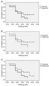
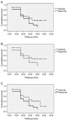
Using Kaplan-Meier survival analysis and comparing the repeat and initial groups survival curves with a log-rank test, we found that the patients who underwent initial trabeculectomy had a greater probability of survival than patients who had undergone previous surgery. But these differences were not statistically significant (p = 0.11 for definition 1, p = 0.14 for definition 2, p = 0.06 for definition 3). According to Kaplan-Meier survival analysis the probability of maintaining an IOP ≤18 mmHg, ≤21 mmHg and 30% decrease from preoperative IOP in repeat group were 25.5%, 32.6%, 17.0%; and in initial group were 47.7%, 52.1%, 45.7% respectively without medications 2 years after surgery (Fig. 1). Estimation of qualified success for definitions 1, 2, 3 showed that 55.8%, 84.4%,and 55.2% of patients in repeat group at 2 years, respectively, versus 60.8%, 73.2% and 54.5% of initial group patients (Fig. 2). Survival curves were compared using the log-rank test and showed no statistically significant difference (p = 0.46, p = 0.92, p = 0.35, respectively).
The early complications observed during the first postoperative month included a shallow or flat anterior chamber in 24 (27.6%) of 87 eyes, hyphema in 8 eyes (9.2%), mild choroidal detachment (confined to anterior to equator) in 11 eyes (12.6%), and early wound leak in 26 eyes (29.9%). Table 6 compares the number and percentage of early postoperative adverse outcomes between initial and repeat surgery groups. None of the eyes either group, developed hypotony maculopathy or endophhalmitis. The number of complications during the follow up period did not differ significantly between the two groups (p = 0.65). Eight eyes in repeat group and 10 eyes in initial group developed more than one complications. One eye in repeat group, and 3 eyes in initial group, had lost two or more lines of Snellen acuity at the last follow up visit compared with their preoperative acuity. In all eyes, it was attributed to cataract progression. For both groups differences between the preoperative and postoperative visual acuity on the Snellen chart were not statistically significant (p = 0.79).
Discussion
The present long-term prospective analysis shows that, the difference between the outcomes of the initial and repeat trabeculectomy is minimal in regard to "qualified success". On the other hand, regardless of the definition, complete success was significantly more common in the initial group and greater proportion of eyes in the repeat group needed postoperative anti-glaucoma medications. Postoperative complication rates were similar in the eyes of two groups.
It has been proposed that previous intraocular surgery is associated with the breakdown of the blood-aqueous barrier, alterations to the composition of growth factors in aqueous and a subsequent greater wound-healing response [21,22]. Moreover, the effect of previous conjunctival surgery other than intraocular one has been shown as a significant risk factor for failure of filtration surgery [9]. It has been shown that the conjunctival changes induced by a previous surgery includes the general increase in subepithelial conjunctival cellularity especially an increase in the number [8]. It can also be suggested that preexisting conjunctival fibrosis may promote further scarring. Moreover conjunctival scarring at the surgical site may cause technical difficulties on subsequent trabeculectomy.
In clinical practice, bleb failure tends to occur early and usually caused by the production, contraction, and remodeling of collagen leading to excessive scarring at the conjunctival episcleral interface. As is well known, the use of intraoperative and postoperative antimetabolites for the manipulation of wound healing and to reducion of the postoperative subconjunctival scarring response improve the outcome of trabeculectomy [20,23-25]. Reported surgical success rates differ from 78% to 84% in high-risk eyes with the use of adjuvant MMC [26,27].
The question of which management strategy is desirable in eyes with failed initial trabeculectomy has long been debated. Postoperative bleb manipulation such as massage, laser suturelysis, removing of releasable sutures, alteration of the tension via adjustable sutures and needling are now considered important parts of postoperative trabeculectomy bleb management [28].
Needling is one of the management strategies in eyes with a failing bleb. Broadway et al. [29] demonstrated that, needlings augmented with 5-FU in the postoperative period, had a mean success rate of nearly 60%. Gutierrez-Ortiz et al. [30] found that the outcome of needling with MMC, was better within 4 months after trabeculectomy. The major limitation of needling procedure is the invisible outline of the flap found in most of the eyes with failed blebs due to excessive scar tissue.
Success rate of surgical reopening of the failed blebs after filtration procedure with antimetabolite augmentation has been reported (maintaining an IOP less 18 mmHg with medications) as 64% [15]. Difficulty of watertight closure and a high incidence of early complications were the major complexity of reopening the failed filter. Another possible disadvantage of reopening the failed trabeculectomy blebs is the difficulty in dissection that may be hampered by bleeding.
Altough, there have been no previous reports which prospectively compares the outcomes of initial and repeat trabeculectomies, glaucoma surgery in an eye with failed initial trabeculectomy is often postponed because of the risk of possible failure and medical therapy is generally preferred. Additionally, repeat surgical treatment is accepted as a technically more difficult procedure. In our study, we had no surgical difficulty in repeat trabeculectomy procedures especially if the first operation had been performed on nasal or temporal superior quadrants. Moreover, there was no statistically significant difference in the number of eyes with complications between the initial and repeat trabeculectomy groups.
Recently Law et al. [31] evaluated retrospectively the outcomes and risk factors of repeat trabeculectomy with MMC as compared with that of initial trabeculectomy with MMC. Investigators reported significantly higher rates of failure for repeat trabeculectomy with more stringent success criteria. With looser IOP reduction criteria the difference between the 2 groups was not found statistically significant. In accordance with the present study, eyes that underwent repeat trabeculectomy required a higher average number of medications than those that underwent initial trabeculectomy.
You et al. [16] reported the results repeat trabeculectomies augmented with subconjunctival or both subconjunctival and subscleral flap application of MMC in eyes with failed initial filtering operation. The total success rates were 86.4%, 90.9%, respectively. Broadway et al. [9] on the other hand, investigated the outcome of repeat trabeculectomy without antimetabolites and found that the surgical success in the re-operated eye was 38%. Their study group consisted of patients with developmental or secondary glaucoma. The use of anti-metabolites in all of our patients are probably responsible for the higher success rate in our study. Cohen et al. [17] in a retrospective study on repeat trabeculectomy with subconjunctival MMC, reported that history of previous surgery did not correlate with postoperative IOP and medications.
One of the major limitations of our study was the modest sample size and follow-up time which may affect the validity of our conclusions and their significance. The durations of glaucoma were significantly different in both groups producing the bias of glaucoma severity on the outcome of trabeculectomy. On the other hand; it is certain that, a longer duration of disease is to be expected if an eye is undergoing a repeat operation. Another limitation of the study is that the criteria for using IOP-lowering medications after trabeculectomy was not standardized.
In conclusion, repeat trabeculectomy augmented with MMC seems to be effective in IOP control. In spite of the need of higher number of anti-glaucoma medications success rates in repeat trabeculectomies with the use of MMC could be comparable with the results of patients who underwent initial trabeculectomy without significant increase in the rate of complications. This information may be useful for planning the surgery in eye with failed bleb and enables the surgeon to counsel the patient before surgery. Results of this study also provide a rationale for the use of antifibrotic agents when performing repeat trabeculectomy.


 XML Download