

 PDF
PDF ePub
ePub Citation
 Print
Print


Keratoconus is a slowly progressive noninflammatory disease of the central cornea which causes central stromal thinning, apical protrusion, and irregular astigmatism. The irregular astigmatism created by ectasia of the central cornea cannot be sufficiently corrected with spectacles, and rigid gas permeable (RGP) contact lenses have become the mainstay of the treatment [1].
Although RGP lenses provide improved vision, they have the potential to damage the cornea. Wearing RGP lenses may be a precipitating factor in the progression of keratoconus in some cases [2]. Scarring of the central cornea may occur in keratoconic eyes not wearing contact lenses; however, it is possible that contact lenses can hasten scarring [1,3]. Several causes may be responsible for the initiation and progression of keratoconus. Keratocyte and stromal alterations, which are associated with an imbalance between proteolytic enzymes and protease inhibitors in the cornea, lead to corneal thinning and scarring [4,5]. Corneal epithelial trauma may be responsible for premature keratocyte apoptosis and may exacerbate stromal changes in keratoconus, resulting in progression [6].
Multicurve RGP lenses have the advantage of minimal contact at the apex because of an increased sagittal height, enabling patients with advanced keratoconus to wear the lens more comfortably. These lenses may also reduce the possible contributions to the progression of keratoconus on a short term basis [7]. Hence, we investigated the effects of wearing multicurve RGP lenses on topographic changes in keratoconus in order to evaluate the contribution of lens use to the progression of keratoconus.
Materials and Methods
Patients
The medical records of all patients who were diagnosed with keratoconus in the contact lens clinic at Seoul National University Hospital between March 1, 2002 and December 31, 2005 were retrospectively reviewed. Keratoconus was diagnosed by positive Rabinowitz indices on topography (Orbscan IIz; Bausch & Lomb, Claremont, CA, USA) and characteristic clinical findings such as apex protrusion or thinning, a Fleischer ring, Vogt's striae, and superficial scarring. One hundred thirty-seven patients were identified. Patients who were prescribed multicurve RGP lenses were included in the lens-wearing group. Keratoconic patients who chose not to wear any type of contact lens were included in the control group. Fifty-six patients who had less than one year of follow-up were excluded. Four patients who had less than eight hours of daily contact lens use and two patients with severe recurrent erosion and discomfort were also excluded.
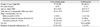
Overall, the lens-wearing group consisted of 77 eyes from 52 patients and the control group consisted of 30 eyes from 23 patients. The mean follow-up periods were 22.6 and 20.5 months in the lens-wearing and control groups, respectively. Demographics of the subjects are summarized in Table 1.
Both the lens-wearing and control groups were divided into a mild to moderate keratoconus group and an advanced keratoconus group; these groups were then analyzed. The stages of keratoconus were classified as mild (central K<45 diopter [D]), moderate (central K between 45 to 52 D), and advanced (central K>52 D) according to the guidelines of the Contact Lens Association of Ophthalmologists [7]. The ocular examination, contact lens fitting, and follow-up examination were performed by the same clinician (MKK).
Lens design and characteristics
The multicurve RGP lens used in this study (YK Lens, Lucid Co., Seoul, Korea) had one base curve and three peripheral curves. It was made from Quantum I (Polymer Technology Corporation, Boston, MA, USA) with a hardness of 84 (Shore D) and an oxygen transmissibility (Dk) of 47 (cm/sec) [(mL O2/mL · mm Hg) · 10-11]. The center thickness was 0.1 mm at -10.0 D and the wetting angle was 48°, as determined by the captive bubble method. The base curves ranged from 5.0 mm to 7.6 mm in 0.1 mm increments, except for the first three curves, which changed in 0.2 mm increments.
Lens fitting method
The 77 eyes in the lens-wearing group were fitted with multicurve RGP lenses based on the topographic index and the examination of fluorescein patterns using slit lamp biomicroscopy. Three-point touch with light apical touch was applied. If there was persistent punctate staining, apical clearance was employed. We followed the guidelines for multicurve RGP lens fitting previously described by Lee and Kim [8]. The initial lens applied to an eye for diagnostic fitting was based on the converted radius, which was derived from the average of the steep and flat keratometric diopteric values. The base curve radius (BCR) was adjusted to accomplish a "light feather touch" at the lens-central corneal interface. The BCR was selected to be 0.1 to 0.15 mm flatter than the BCR that first demonstrated definite apical clearance. The radii of the peripheral curves were modified to establish three-point contact, to allow for interchange of mid-peripheral tears and to achieve an adequate axial edge lift of approximately 0.2 to 0.25 mm. The optic zone diameter was changed in 0.5 mm increments in accordance with BCR changes; 5.0 mm in BCR ranged from 5.0 to 5.9 mm, 5.5 mm in BCR ranged from 6.0 to 6.5 mm, 6.0 mm in BCR ranged from 6.6 to 6.9 mm, and 6.5 mm in BCR was 7.0 mm or flatter. The total diameters of the prescribed lenses ranged from 8.6 mm to 8.8 mm. If the first lenses were not acceptable, the lenses were reordered with appropriate changes within three weeks after the first fitting.
Evaluation
Changes in topographic indices were evaluated before multicurve RGP lens fitting at the baseline visit and at the final visit using Orbscan IIz. Topographic evaluations were performed one hour after multicurve RGP lens removal. During follow-up, patients were asked about their average daily uses and overall comfort levels. The best corrected visual acuities were evaluated in both the lens-wearing and control groups. Slit lamp biomicroscopic examination was performed to determine the presence of corneal scarring and temporary or persistent punctate corneal staining (Efron's grading scale) [9]. Punctate staining that disappeared spontaneously or with treatment by the next consecutive visit was defined as "temporary punctate staining." If the fit of the lens was acceptable, preservative-free lubricant eyedrops were prescribed for temporary punctate staining. If not, the lenses were refitted stepwise until the staining decreased. Staining at more than two consecutive visits and which did not disappear despite use of the lubrication drops was defined as "persistent punctate staining." In these cases, the fitting method was changed to account for apical clearance.
To evaluate changes in the topographic indices possibly due to lens wearing, several parameters including Sim Kmax, Sim Kmin, apical power (the power of the cone apex), anterior and posterior elevations, thinnest corneal thickness, and the irregularity index at 3 mm (IR) were compared between the lens-wearing and control groups during follow-up. The usefulness of the Orbscan system in detecting keratoconus has been reported [10-15]. Although the reproducibility of the Orbscan in the evaluation of keratoconus still remains a matter of controversy, its parameters, including anterior and posterior elevations, have been shown to be clinically relevant for detecting keratoconus and suspected keratoconus [16-19]. The topographic index used in the analysis of keratoconus progression was obtained from staining-free or healed corneas. The differences in anterior and posterior elevations were examined in each group using the differential map from the Orbscan IIz. The acoustic factor for pachymetric measurement was set to its factory default value of 0.92. In keratoconus patients, Orbscan IIz is a valid clinical tool for the noninvasive assessment of corneal thickness [20].
Intra-group comparisons were performed to evaluate topographic changes during follow-up using the paired t-test, and inter-group comparisons were performed using the independent t-test. Statistical analysis was performed on a personal computer using the SPSS ver. 12.0 (SPSS Inc., Chicago, IL, USA). The level of statistical significance was set at a p-value of 0.05.
Results
The logarithm of the minimum angle of resolution (logMAR) visual acuities of the lens-wearing and control groups at the first visit were 0.68±0.61 and 0.54±0.47 with best spectacle correction, respectively (p=0.255). In the lens-wearing group, visual acuity significantly improved to -0.016±0.065 (p<0.001) after multicurve RGP lens fitting. At the end of the follow-up period, the multicurve RGP lens-corrected visual acuity of the lens-wearing group improved to -0.032±0.10 (p=0.05, paired t-test). The multicurve RGP lenses were reported to be comfortable in 67 eyes (87.0%), slightly uncomfortable in seven eyes (9.1%), and uncomfortable in three eyes (3.9%). The mean daily use was 11.6±3.0 hours.
The incidences of punctate corneal staining, including transient and persistent erosions, in the lens-wearing and control groups were 32 eyes (41.6%) and two eyes (6.6%), respectively, illustrating a significantly higher incidence in the lens-wearing group (p<0.001). Corneal scarring was newly detected during follow-up in three eyes (3.9%) and two eyes (6.7%) in the lens-wearing and control groups, respectively.
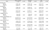
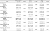
In the lens-wearing group with advanced keratoconus, the Sim Kmax, Sim Kmin, apical power, astigmatic index, and anterior elevation significantly decreased during the follow-up period (Table 2). The thinnest corneal thickness and irregularity index decreased, and the posterior elevation increased in the lens-wearing group, but these changes were not significant. In the lens-wearing group with mild to moderate keratoconus, significant changes were not found, except for posterior elevation. In the control group, the apical power and IR increased from 55.56±7.25 D to 57.11±7.75 D and from 3.06±1.68 D to 3.25±1.71 D, respectively (p=0.008, p=0.01) (Table 3).
We compared changes in the topographic indices from baseline to the final visit between patients in the lens-wearing group who demonstrated punctate staining and patients in the lens-wearing group who did not demonstrate punctate staining. In the lens-wearing group with punctate staining, significant changes were not found, except for Sim Kmax, which decreased from 54.01±5.85 D to 52.83±5.38 D (p=0.045). In the lens-wearing group without punctate staining, the Sim Kmax, Sim Kmin, and apical power decreased. The Sim Kmax decreased from 54.40±5.07 D to 53.28±3.32 D, the Sim Kmin from 48.63±3.32 D to 47.95±3.32 D, and the apical power decreased from 60.76±6.30 D to 58.33±5.51 D (p=0.032, p=0.05, p=0.001, respectively). The IR did not significantly change in either group.
Discussion
The adequate fitting method for RGP lenses in patients with keratoconus includes apical bearing fitting with primary lens support on the apex of the cornea, apical clearance fitting with lens support and the bearing directed off the apex and onto the paracentral cornea, and three-point touch fitting with central light touch and mid-peripheral touch with peripheral clearance [3,21,22]. Apical bearing fitting may induce chronic apical epitheliopathy, anomalous apoptosis, loss of rigidity, and thinning [23]. Apical clearance fitting may induce an increase in curvature and promote ectasia [24]. Considering that most of the lens-bearing pressure is supported by the thicker peripheral cornea and that light central touch does not cause significant staining, the preferred technique is the three-point touch [23,24].
Contact lens wear itself may cause progression of keratoconus, regardless of the fitting method. A progressive increase in the corneal curvature might develop if the contact lens fitting is inadequate for the cone [25]. In fact, we observed an increase in distortion with excessive apical bearing fitting or apical clearance fitting in two of our patients (unpublished data). This is the reason why we investigated the topographic changes after lens use in patients with keratoconus, and we indeed found that the Sim Kmax, Sim Kmin, astigmatic index, apical power, and anterior elevation all significantly decreased in the lens-wearing advanced keratoconic eyes over an average period of 23.2 months. More importantly, the IR did not change in the lens-wearing group; on the contrary, IR significantly increased in the control group. Hence, it is not likely that wearing multicurve lenses with minimal apical touch fitting contributed to progression of keratoconus, even though they may result in transient punctate staining. Topographic indices are important in determining the severity and progression of keratoconus; however, they are sometimes limited due to poor reflex images. Increases in the central corneal curvature, refractive power, and corneal irregularity with widening of the cone are risk factors for the progression of keratoconus [1,25,26]. However, to confirm the progression, other clinical data such as corneal scarring, corneal signs, and spectacle visual acuity are required [27]. We did not observe any clinically definitive evidence of progression because almost none of the patients exhibited clinical signs, such as severe scarring. Nevertheless, the follow-up period of approximately 22 months is too short to conclusively determine the effect of lens wear on the progression of keratoconus; we still believe these results are worthy of note because many keratoconic patients continue to wear contact lenses.
Epithelial trauma may be associated with the pathogenesis or the progression of keratoconus through the development of stromal thinning [28]. Persistent epithelial damage can produce inflammatory cytokines and degrading enzymes, resulting in apoptosis of keratocytes and stromal thinning. Depending on the degree of contact, apical support fittings, including the three-point touch, are likely to induce epithelial trauma in the cone apex compared with apical clearance fitting [23]. We adopted a three-point touch fitting method for use in our patients and found that the incidence of temporary punctate corneal staining in the lens-wearing group was higher than that of the control group, although there was no significant difference in the incidences of persistent punctate corneal staining and corneal scarring (three eyes [3.9%] vs. one eye [3%], three eyes [3.9%] vs. two eyes [6.7%], respectively). Only three eyes were fitted using the apical clearance method. These eyes were initially fitted with a three-point touch; however, they were refitted using the apical clearance method due to persistent punctate staining. We evaluated changes in the topographic indices from baseline to the final follow-up visit in eyes with a history of punctate corneal staining and in those without corneal staining; the topographic indices did not show any significant changes in eyes with punctate staining. Accordingly, temporary punctate staining caused by light central touch in three-point touch fitting does not appear to cause increases in curvature or irregularity in keratoconus, at least during a medium length follow-up period.
To confirm whether the regression effect is irreversible or temporary, lens wearing should ideally be avoided for at least three months, and the topographic indices should be re-evaluated. However, the cessation of lens use is not ethically, since poor visual acuity would be intolerable. In the present study, all topographic evaluations were performed one hour after lens removal. Although our evaluation did not include full cessation of contact lens use prior to examination, we believe that it still provides useful information for understanding the likelihood of progression of keratoconus with contact lens use.
Another important factor to be considered when assessing keratoconus progression is age. Longitudinal studies have shown a flat K slope in patients younger than 35 years (0.41±1.1 D), greater than that in patients older than 35 years (slope, 0.1±0.4 D/yr; p<0.0001) [29]. In our study, the mean ages of the lens-wearing and control groups were not significantly different, with both being less than 35 years of age. Therefore, our study on the progression of keratoconus does not appear to be influenced by age. Another consideration regarding the progression of keratoconus is corneal thickness. We found a decrease in corneal thickness in the lens-wearing group; however, this finding was not statistically significant. Therefore, long-term investigation is now needed to evaluate lens-wearing effects on corneal thickness in patients with keratoconus.
RGP fitting with multicurve lenses is not likely to contribute to progression of keratoconus. However, a long-term longitudinal study investigating the long-term effects of RGP fitting is needed.
 XML Download