

 PDF
PDF ePub
ePub Citation
 Print
Print


In cases of monocular vision, many patients have a large horizontal deviation with or without vertical deviation [1,2]. Even though bilateral surgery on multiple extraocular muscles may effectively correct these deviations, patients and surgeons alike are hesitant to proceed with surgery on the only normal eye.
We effectively corrected a patient with large exotropia and considerable hypertropia using unilateral recession-resection surgery of the horizontal recti muscles with inferior displacement and augmented anterior transposition of the inferior oblique (IO) muscle with a posterior intermuscular suture.
Case Report
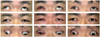
A 50-year-old male patient had a history of exposure to a bomb explosion 22 years previously. He had no significant medical or ophthalmic history prior to the trauma. An examination revealed visual acuities of no light perception in his right eye and 20/20 in his left eye. A pale and atrophic optic nerve head was observed in the right eye. He had right exotropia of 80 prism diopters (PD) and right hypertropia of 20 PD (Fig. 1, left).
All surgical procedures were performed undertopical anesthesia with 0.5% proparacaine hydrochloride ophthalmic solution. A forced duction test was unrestricted. A recession-resection procedure of the horizontal recti muscles with inferior displacement was planned for the exotropic eye. Medial rectus resection of 6.0 mm was performed with 4.0 mm of inferior displacement from the original insertion site. Following this, the lateral rectus (LR) muscle was carefully isolated. We initially wanted to perform an LR recession of 12.0 mm with 4.0 mm of inferior displacement. However, the IO muscle interfered with reattachment of the LR muscle,and we subsequently decided to sever the IO muscle.
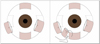
While the inferior rectus (IR) muscle was secured with a muscle hook, the IO muscle was isolated and severed. The LR muscle was reattached 12.0 mm posterior and 4.0 mm inferior to the original insertion site (Fig. 2, left). The prism cover test was then performed using the Krimsky method. Approximately 10 PD of exotropia remained, and a slight limitation of motion during infraduction was noted; however, there was no definite hypertropia.
Finally, the anterior edge of the previously disinserted IO muscle was placed just lateral to the IR insertion. The posterior edge of the IO muscle was sutured 2.0 mm anterior to an imaginary line extending from the IR insertion. The posterior intermuscular augmentation suture was placed at 6.0 mm posterior to the muscle insertion (Fig. 2, right) [3]. The prism cover test was repeated,and approximately 10 PD of exotropia still remained. Although a slight limitation of motion during infraduction was still noted, there remained no definite hypertropia.
One week later the patient was found to have 10 PD of right exotropia in the primary position and a slight limitation of motion during infraduction (Fig. 1, middle). After one month, 30 PD of right exotropia had re-developed; however, this remained stable at five months postoperative (Fig. 1, right) and no additional procedure was necessary.
Discussion
Many patients with monocular vision have a large deviation of the blind eye [1,2]. Even though bilateral surgery on multiple extraocular muscles may effectively correct these deviations, patients and surgeons alike are hesitant to proceed with surgery on the only normal eye.
In the present case, the patient had large right exotropia combined with hypertropia. He strongly refused surgery on his left eye so we proceeded with unilateral recession-resection surgery with inferior displacement of the horizontal recti muscles. However, the position of the IO interfered with a recessed and inferior-displaced attachment of the LR. The IO was disinserted, and the LR was subsequently attached at the desired position. We then performed an IO anterior transposition augmented with a posterior intermuscular suture.
Although anterior transposition of the IO muscle was not a planned procedure, it resulted in the IO serving not only as an anti-elevator, but also as a depressor. This arrangement might therefore be effective in treating hypertropia and limited infraduction. Postoperatively, supraduction was slightly limited (Fig. 1, right)and seemed to originate from the IO anterior transposition. However, it did not result in any cosmetic problems, and thepatient was very satisfied. Other surgical options could have certainly been performed, including-recession-resection surgery of the horizontal recti muscles with inferior displacement orrecession-resection surgery of the horizontal recti muscles combined with superior rectus recession.However, these surgical options have serious risks, including anterior ischemic syndrome. Although the patient's ocular alignment was not perfect at five months postoperative, our procedure was easy and safe.
Posterior augmentation intermuscular suturing was first employed in 1995 by Buckley and associates for full tendon transposition in paralytic strabismus [3,4]. We previously gained experience with recession-resection surgery combined with augmented full tendon transposition with a posterior intermuscular suture in a patient with a large horizontal and vertical deviation [5].
There are several possible causes for the intraoperative finding of limited infraduction following the recession-resection procedure. While we performed all procedures under topical anesthesia, the eyeball may not have been able to freely move intraoperatively because of edema in the adjacent tissue that may have interfered with ocularmovement. In addition, the previous traumatic changes may have had some unexpected influence.
In conclusion, we corrected large exotropia combined with hypertropia by means of a single unilateral surgery. Unilateral recession-resection surgery with inferior displacement and augmented anterior transposition of the IO with a posterior intermuscular suture can effectively treat a patient with large exotropia and considerable hypertropia.
 XML Download