

 PDF
PDF ePub
ePub Citation
 Print
Print


Uveitis is a complex intraocular inflammatory process primarily involving the uvea, retina, and other intraocular structures. This intraocular inflammation can be either acute or chronic and is readily induced in laboratory animals by sensitization with the retinal soluble protein S-antigen. The resultant inflammation is known as experimental autoimmune uveo-retinitis (EAU).1 Previously, in EAU, we quantified the nitric oxide (˙NO) production by the Griess reagent, and we and others evaluated the inducible nitric oxide synthase (iNOS) expression as the means of ˙NO production in inflammation.2-6 Although quantification of nitrite, nitrate, nitrosylhemoglobin, and methemoglobin generally reflects ˙NO production in the biological system, interference in these assays by the tissue components is well known. Nitric oxide synthase presumably takes L-arginine as a substrate, converting it to ˙NO and L-citrullin. However, in recent years, this direct link between NOS and ˙NO was somewhat weakened by several new findings: 1) Nitric oxide synthase does not directly synthesize ˙NO; rather, it synthesizes nitroxyl anion (NO-), which can subsequently be converted to ˙NO in the presence of superoxide dismutase (SOD);7 and 2) The quantity of NOS present does not directly reflect the amount of ˙NO produced.8 Therefore, a direct means of establishing the generation of ˙NO in EAU is necessary.
Nitric oxide is a paramagnetic species that binds with high affinity to a variety of metal chelates. Therefore, electron spin resonance (ESR) spectroscopy with spin trapping reveals a direct evidence of ˙NO presence. ESR can also specifically detect ˙NO and distinguish it from three other redox forms. Two dithiocarbamate derivatives have frequently been used in the trapping of ˙NO in living tissues.9-11 These are ferrous iron complexes of N-methyl-D-glucamine dithiocarbamate, (MGD)2-Fe2+, and diethyldithiocarbamate, (DETC)2-Fe2+. The high binding constant and water solubility of (MGD)2-Fe2+ and the stability of the corresponding adduct [(MGD)2-Fe2+-NO] have made it possible to detect low concentrations of NO formed in biological systems.12-14
Chemiluminescence is a photon emission process that is specifically enhanced by the presence of 5-amino-2,3-dihydro-1,4-phthalazinedione (luminol).15,16 It has been widely used in the past in the detection of reactive radical species such as O2-, hydrogen peroxide, and the hydroxyl radicals released from cells and tissues.15-19
In vitro, the ˙NO and O2- radicals are known to combine to form ONOO-; the rate of the combination reaction is limited only by diffusion (K=3.7×10⁷ M/s).20 Peroxynitrite formation in vivo, however, is more difficult to detect, since ONOO- is protonated with decomposition at physiological pH. It has recently been shown that in alkaline conditions (pH>9), the half-life of ONOO- is considerably prolonged.20 Therefore, this type of stabilization could facilitate the detection of ONOO- generated in vivo. It has been reported that ONOO- can induce luminol chemiluminescence that is inhibited by SOD or urate. It has further been shown that the bicarbonate/luminol system can specifically discern the presence of ONOO- in the pool of reactive species by the formation of a labile intermediate ONOOC(O)O between ONOO- and bicarbonate. This bicarbonate luminol adduct then decomposes to the active radical species, increasing the quantum yield to at least 2-fold from that of ONOO- without bicarbonate.21 Since no other reactive species released by the inflammatory phagocytes display this phenomenon, the extent of this increase signifies the presence of ONOO- in the active radical pool.
In this study, we used ESR spin trapping with the (MGD)2-Fe2+ complex to unequivocally detect the synthesis of ˙NO in the EAU retina and choroid for the first time. In addition to establishing the g-values and hyperfine splittings of the EAU samples, several controls with or without a spin trap were evaluated. An authentic [(MGD)2-Fe2+-NO] complex was also prepared chemically to aid in the characterization of this adduct in the EAU retina. We have also quantified the photon-emitting species generated at the inflammatory site by luminol chemiluminescence. Bicarbonate was introduced in these systems, and the increase caused by this addition was quantified to demonstrate for the first time that the luminescent reactive species were mostly ONOO-.
Materials and Methods
Induction of EAU
The uveitogenic human S-antigen peptide, DTNLASSTIIKEGIDRTVLG, was synthesized on 4-hydroxy-methylphenoxymethyl resin using an automated peptide synthesizer (model 430A; Applied Biosystems, Foster City, CA) and desalted on a Sephadex G-10 column (Sigma, St. Louis, MO).1 The purity was assessed by reversed phase high performance liquid chromatography (Bio-Rad, Richmond, CA). Male Lewis rats, each weighing 150-175 g (VAF, Charles River Laboratory, Wilmington, MA), were given a hind foot-pad with 100 ㎍ of human S-antigen peptide in Freund's complete adjuvant to induce EAU, as previously described. At the time of immunization, each animal also received an intravenous injection of 1 ㎍ pertussis toxin (List Biological Laboratory, Campbell, CA) in 0.3 ml sterile saline. A total of 117 rats were immunized; another 12 were used as non-immunized controls. Immunized animals were sacrificed at the peak of inflammation, at between 12 and 13 days post-immunized. The eyes were enucleated, the anterior segments were removed, and the retina and choroid were collected. All procedures conformed to the Association for Research in Vision and Ophthalmology Resolution on the Use of Animals in Research.
Electron spin resonance spectroscopy and spin trapping
All procedures involving a spin trap were conducted under dim light. The spin trap complex (MGD)2-Fe2+ was prepared in 0.01 M phosphate-buffered saline (PBS) with final concentrations of 13.5 mM FeSO4.7H2O and 86.0 mM MGD (Oxis, Portland, OR). Two 10 ㎕ doses each of (MGD)2-Fe2+ solution were injected into the vitreous cavity at different scleral sites. Similarly, two 10 ㎕ injections containing 0.01 M PBS only were made in the control animals. Ninety minutes after injection, the animals were sacrificed, the globes were enucleated, and the retina and choroids were removed. The retina and choroid from two eyes were combined and homogenized briefly (two seconds) in 0.4 ml of (MGD)2-Fe2+ solution before the mixture was transferred into the ESR tube (Wilmad, Buena, NJ). The tube was incubated at room temperature for 30 minutes, and the ESR measurement commenced immediately at the end of incubation. For the inhibition experiments with AG, the globes were injected intravitreally with two 10 ㎕ doses of 12 mM AG at different sites, two hours prior to injection of the spin trap (MGD)2-Fe2+, as described above. An authentic (MGD)2-Fe2+-NO complex was prepared chemically. Aliquots of the MGD and FeSO4·7H2O stock solution were mixed to yield final concentrations of 6 mM MGD and incubated with 10 ㎛ of the ˙NO donor, S-nitroso-N-acetyl-penicillamine (SNAP; Cayman Chemical Company, Ann Arbor, MI); the reaction product was transferred into the ESR tube.
Electron spin resonance spectra were recorded at 77 K using a Varian E-109 Spectrometer (Varian Instruments, Palo Alto, CA) equipped with a TE-102 cavity and a cold-finger dewar. Other instrument settings were as follows: 20 mW microwave power, 9.28 GHz frequency, 4×10⁴ gain, 100 KHz modulation frequency, 4 G modulation amplitude, 128 ms time constant, and 2 minute scan time. Scanning was repeated at least four times. All ESR measurements were carried out using a combination of two eyes as one sample, and each measurement was repeated at least three or four times, using a total of six to eight eyes for each category of measurement.
Measurement of luminol chemiluminescence
A stock solution of luminol (Sigma, St. Louis, MO) was prepared by first dissolving 1 mg of luminol in 100 ㎕ of dimethyl sulfoxide and then diluting it to 10 ml with 0.01 M PBS (pH 7.4). Exactly 400 ㎕ of the stock solution was diluted to 200 ml with 0.01 M PBS to provide the working solution. The final concentration of luminol in the working solution was 0.2 ㎍/ml. The final concentration of dimethyl sulfoxide was 0.28 mM. All procedures were conducted under dark illumination.
Each sample, consisting of retinas and choroids collected from six eyes, was placed in a polypropylene counting vial, and 10 ml of luminol working solution was added immediately before counting. Chemiluminescence was counted on a Packard liquid scintillation analyzer (Packard Instruments, Downers Grove, IL) with the operating mode set on single photon counting. This mode sets the two photomultiplier tubes in out-of-coincidence counting, thereby maximizing the counting efficiency for weak photon emission. All scintillation vials and solutions used were dark-adapted overnight prior to use to reduce background counting. Samples were counted for one minute immediately after mixing with luminol. The counting was then repeated for several cycles until the counts were stabilized. For the bicarbonate and inhibitor experiments, the retina/choroid samples were first counted until maximal stable counts were established, and then sodium bicarbonate (50 mM final concentration), SOD (5,270 units; Sigma), N-nitro-L-arginine methyl ester (L-NAME, 1 mM final concentration, Sigma), and AG (5 mM final concentration, Sigma) were added. In these experiments, counts without the inhibitor were first established by counting for 15 to 20 minutes; the inhibitor was then introduced, and the counting resumed for an additional 50 to 60 minutes. Alternatively, a parallel counting procedure was employed. Briefly, two vials with the same amount of tissue were counted for 10 to 15 minutes, bicarbonate/inhibitor was added to one of the vial, and counting for both vials was resumed for 50 to 60 minutes thereafter. These two counting procedures yield similar results. Each experiment was repeated at least three times, using a total of 18 globes for each category of experiment.
Results
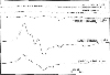
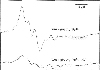
Following an intravitreal injection of the freshly prepared spin trap (MGD)2-Fe2+ into the eyes of the EAU rats, a prominent three-line ESR spectrum (aN=12.5 G; giso=2.04) was obtained (Fig. 1). The spectrum unambiguously indicated the adduct [(MGD)2-Fe2+-NO] and was consistent with that reported in the postischemic heart.22,23 There was no appreciable signal in either the normal controls or the EAU rats injected with 0.01 M PBS without the spin trap (Fig. 1). Control animals injected with the same concentration of (MGD)2-Fe2+ yielded a group of low intensity multicomponent signals that are not characteristic of any known radical species (Fig. 1). In this spectrum, one of the more obvious broad signals from control animals with the spin trap is unrelated to the [(MGD)2-Fe2+-NO] triplet, and its identity is not known. This particular signal appears to be associated with the spin trap and some component of the naÏve retina, irrespective of the development of uveitis, since the chemically synthesized retina-free [(MGD)2-Fe2+-NO] adduct resulted in no such signal. In the inhibition experiment, AG, which is an iNOS specific inhibitor, considerably suppressed the triplet signal (Fig. 2); the intensity of the [(MGD)2-Fe2+-NO] signal was reduced to one-tenth or less of the corresponding signal without the inhibitor. In this experiment, AG was injected two hours before the injection of the spin trap. Aminoguanidine injected either three or four hours before administration of the spin trap resulted in further suppression (data not shown). The authentic [(MGD)2-Fe2+-NO] prepared from the ˙NO donor, SNAP, and the spin trap was also used in generation of an ˙NO signal. With the concentration (10 µ M) of SNAP used, the triplet signal generated was not as intense as those seen in EAU tissues; nevertheless, the central g-value and hyperfine splitting are indicative of the [(MGD)2-Fe2+-NO] adduct and similar to those obtained from EAU eyes plus spin trap.
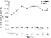
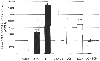
Luminol chemiluminescence was measured with the liquid scintillation analyzer set at out-of coincidence photon counting to maximize the counting efficiency. The chemiluminescence counting from a pool of six sets of retina and choroid at the peak of inflammation were on the order of 70,000 counts. The counts usually started immediately following the addition of luminol, reached a plateau in 20 minutes, and then decayed slowly after 40 minutes. A high level of counting persisted even after 60 minutes. At the plateau, the phagocytic chemiluminescence was at least six-fold higher than those derived from the non-inflammatory source (Fig. 3). The maximum counts obtainable for a specific inflammatory model appear to be highly dependent on the degree of inflammation. In the luminol chemiluminescence, after stabilization of the count, the exogenous addition of bicarbonate increased the maximal counts by more than two-fold and sustained this level for two hours thereafter. Without bicarbonate, the counts decayed after one hour. The enhancement in quantum yield by the introduction of bicarbonate has been attributed to the presence of ONOO- in the system and the subsequent formation of the labile intermediate ONOOC(O)O- to facilitate an increase in radical generation.21 Using bicarbonate/luminol chemiluminescence in the inflamed retina, we evaluated the combined effects of the inhibitors SOD (5,270 units), L-NAME (1 mM), AG (5 mM), and AG (5 mM) + SOD (5,270 units). In these experiments, the counts without the inhibitors were first established, the desired amount of inhibitor was then introduced, and counting was resumed for 60 minutes thereafter. The extent of inhibition displayed with a particular inhibitor was subsequently confirmed with a second procedure. Two vials, one with and one without inhibitor, were counted in parallel. In some instances, the addition of the inhibitor caused a temporary surge in the counts; however, these counts rapidly returned to their respective levels, where they remained for at least 40 to 50 minutes. The two procedures yielded similar degrees of suppression. Using these procedures, 1 mM of L-NAME, an inhibitor of both constitutive and inducible NOS, was found to give 22% suppression. With either 0.1 or 2 mM concentrations of L-NAME, the change in the suppression level was found to be insignificant (data not shown). The inhibition was increased to 37% with 5 mM of AG, while SOD inhibited 40% of the chemiluminescent activity. In contrast with SOD, chemiluminescence was inhibited by 40%. A marked additive effect of 77% inhibition was seen with the combination of AG and SOD (Fig. 4).
Discussion
In the present study, we obtained direct evidence of ˙NO generation in the retina and choroid of EAU animals. The production of ˙NO was unequivocally determined by ESR, using (MGD)2-Fe2+ complex as a ˙NO trap. The three-line spectrum (central g value of 2.04 and hyperfine splitting of 12.5 G) obtained was characteristic of the adduct [(MGD)2-Fe2+-NO]. The majority of this signal was eliminated by the AG intravitreally injected into the eye, as AG is a specific blocker for iNOS. In the inflamed retina, we also detected an approximately six-fold increase in chemiluminescent activity in comparison to those from the non-inflamed controls. These luminescent counts were further increased more than two-fold by the addition of bicarbonate to the isolated retina and choroid ex vivo, a phenomenon accounted for by the presence of a high steady-state concentration of ONOO-. Further, this luminescent activity was significantly inhibited by the addition of either AG or SOD separately, and markedly inhibited by the addition of AG and SOD combined. Again, this suggests that both ˙NO and O2 participate in the formation of the major chemiluminescent species in the inflamed retina, namely ONOO-. Neither ˙NO nor O2 alone was capable of directly producing luminol chemiluminescence.
Nitric oxide binds to (MGD)2-Fe2+ and forms the paramagnetic d7 state mononitrosyl iron complex [(MGD)2-Fe2+-NO], yielding the characteristic triplet ESR spectrum.24 Thus, (MGD)2-Fe2+ is selective in trapping ˙NO, since the trapping of ˙NO, one of the redox forms of nitrogen monoxide, will result in a diamagnetic low spin D8 state undetectable by ESR spectroscopy. In the trapping of ˙NO at the peak of inflammation, we obtained a prominent triplet signal of the [(MGD)2-Fe2+-NO] adduct 10 times higher in intensity than that obtained from non-immunized animals injected with the same spin trap. Although the symmetry of the triplet in the ex vivo system is often not as distinct as those from the in vitro system [see, for example, ref. 24], the g-values and hyperfine splittings obtained are comparable to those reported in tissues for the same radical.25,26 We have also obtained a signal from an authentic adduct, prepared independently from (MGD)2-Fe2+ and the ˙NO donor SNAP; the spectrum and the g-value were very much in agreement with those from the EAU retinas. Little or no NO production is derived from injected (MGD)2-Fe2+, as shown in control rats injected with (MGD)2-Fe2+. This signal was also absent in EAU rats without the spin trap, indicating efficient trapping by (MGD)2-Fe2+ and subsequent stabilization and accumulation of (MGD)2-Fe2+-NO in the retina.
In this experiment, intravitreally injected AG caused nearly complete suppression of the [(MGD)2-Fe2+-NO] signal in the retina. Aminoguanidine, a nucleophilic hydrazine compound, is a known selective inhibitor of iNOS. The inhibitory action is believed to act on the enzymatic activity of iNOS.27 Therefore, the marked suppressive effect of AG injected 3.5 hours prior to the sacrifice of the animal implies: first, that a large proportion of ˙NO synthesis is L-arginine/iNOS-mediated, and second, that there is a relatively fast turnover of ˙NO in the retina, so that the depletion of endogenous iNOS for at least 3.5 hours causes a significant reduction in the retinal ˙NO concentration, as indicated by ESR spin trapping. In the presence of O2 at the site of ˙NO generation, the major route of ˙NO disappearance is by a fast combination reaction to yield ONOO-. In the absence of O2, ˙NO is oxidized and disproportionated to yield nitrite and nitrate, which accumulate as stable products in tissues. With the concentrations injected into the eye for the present study, the (MGD)2-Fe2+ complex appears to compete favorably with other fates of ˙NO in the retina, since the adduct [(MGD)2-Fe2+-NO] obtained was in relatively high concentrations.
The (MGD)2-Fe2+ complex was delivered to the retina both by intravitreal injections to the eyes of live animals and by subsequent incubation of the isolated retinas with the (MGD)2-Fe2+ complex. Spin trapping of endogenously generated radicals within the retina has never been attempted in the past. The concentrations of (MGD)2-Fe2+ used in the present study were comparable to those reported for other tissues.9,11,14 These (MGD)2-Fe2+ concentrations have been shown to pose no deleterious effects.9,11,28 During the course of spin trapping, the free iron was unlikely to be present in the retina, since there is an excess amount of MGD present for chelating exogenous iron (MGD:iron ratio=5:1). Therefore, it is safe to conclude that intravitreal injections of (MGD2-Fe2+ complex will not lead to any deleterious reactions, including the stimulation of ˙NO production. This assumption was subsequently verified by the experiment in which control animals injected with the same concentration of (MGD)2-Fe2+ gave no significant [(MGD)2-Fe2+-NO] signal (Fig. 1).
Chemiluminescence counts for the phagocytic activity in inflammation appear to be highly system-dependent, possibly because of differences in the extent of inflammation and the size of tissues used. The reported maximal counts have been highly variable.19 In this study, the chemiluminescence counts usually displayed a plateau at 20 to 40 minutes after the addition of luminol and decreased thereafter. The addition of bicarbonate to the inflamed retina rapidly increased the counts to more than two-fold. The half-life of ONOO- was prolonged in the alkaline solution, since the protonation/decomposition reaction was largely eliminated. Therefore, the addition of bicarbonate stabilizes ONOO-, thus facilitating the detection of ONOO- in tissues. Moreover, ONOO- appears to be the major species in the pool of reactive radical species, since the two-fold enhancement obtained by adding the bicarbonate is similar to the in vitro observations reported with the pure ONOO- preparation.21
The exact pathway by which bicarbonate enhances the ONOO- luminescence is not known. The light-emitting excited species is apparently produced by ONOO- and HCO3- interactions. Although several thermodynamically feasible speculations have been advanced, the exact nature of this excited species is yet to be proved.29 One fate for this unstable intermediate is to proceed to a facile decomposition that yields O2-.21 Since the inhibitory effect of SOD (Fig. 4) indicates the participation of O2- in chemiexcitation, the increased production of O2- could, therefore, translate to a higher photon quantum yield.
The results of the bicarbonate luminol chemiluminescence indicate that although ONOO- is short-lived at physiological pH (t1/2<1 s), the inflammatory infiltrates are viable for a period of hours after the sacrifice of animals. During this period, these inflammatory cells were continuously activated to express NOS and NADPH oxidase and to produce NO and O2-. Superoxide is known to participate not by directly reacting to luminol, but rather by reacting with the luminol radical formed by the one-electron oxidation of luminol by oxidants, such as the OH˙ radical, lipid hydroperoxide, or other species with similar reactivity.17 Therefore, the observed inhibitory effect of SOD can arise from the inhibition of ONOO- formation from O2- and ˙NO, or from the reaction of O2- with luminol radicals. In this context, the less than 50% inhibition exhibited by SOD might reflect the accessibility of SOD to the O2--generating site in the retina within the short time frame between SOD addition and measurement of the photon emission.
The exogenous addition of L-NAME or AG inhibits the function of NOS. Since AG is a specific inhibitor of iNOS and L-NAME is an inhibitor of both inducible and constitutive NOS, the larger suppression seen by AG might imply that iNOS is the predominant form in the production of ˙NO in this system. However, even the suppression seen with AG is less than 50%. In this experiment, AG was added immediately before the addition of luminol and the measurement of chemiluminescence. Therefore, only the amount of ˙NO newly synthesized within this time frame will be affected. The suppression, however, was more remarkable when instantaneous scavenging of O2- by SOD was added to this iNOS blocker.
Bicarbonate anion is an abundant constituent of the extracellular milieu, present at concentrations as high as 25 mM. Therefore, in biological systems, bicarbonate anion interacts rapidly with ONOO- to form ONOOC(O)O- and enhances the toxicity of ONOO-, as seen in bicarbonate luminol chemiluminescence. Based on kinetic considerations, the HCO3-/CO pair should compete favorably with other biological targets, such as sulfhydryls, for reaction with ONOO- in the extracellular space.29 Heterolytic cleavage of ONOOC(O)O- will also give rise to a potent nitrating agent that will regenerate HCO3-. This reaction is consistent with the catalytic effect of bicarbonate on ONOO--dependent nitration of aromatics, including tyrosine, reported previously.30,31
In summary, we have demonstrated the production of ˙NO in EAU by ESR spin trapping. This method surpasses any other method for indirectly detecting the presence of ˙NO species in the tissue. This reactive nitrogen species then forms the potent biological oxidant ONOO- in the inflamed retina and choroid. Recognition of the species responsible for the tissue injury in EAU will aid in developing therapeutic interventions for the prevalent intraocular inflammations.
 XML Download