

 PDF
PDF ePub
ePub Citation
 Print
Print


The term coloboma refers to "a notch, hole, or fissure in any ocular structure from a congenital malformation or an acquired process".1 Eyes with congenital colobomata and cataracts are at greater risk for complications during cataract surgery because of ocular malformations resulting from embryological maldevelopment.2 Given the abnormal development of the sclera, uvea, and zonules, the structural integrity of the globe may be compromised, increasing the risk of vitreous loss during cataract extraction.
The development of phacoemulsification, and continuous curvilinear capsulorhexis (CCC) techniques have made it possible to improve cataract surgery outcomes. However, visual function may not become normal after surgery due to iris malformation and zonular defects. Nevertheless, the insertion of a capsular tension ring (CTR) into the capsular bag to maintain its circular contour has improved in both visual outcomes and surgical safety. The use of CTRs has been proven to be beneficial for IOL centration.3 When a zonular defect is present and a CTR is inserted at any stage of the procedure to reestablish the capsule's contour, the CTR protects against capsular fornix aspiration, consecutive zonular dialysis extension, irrigation fluid flow behind the capsule, vitreous herniation into the anterior chamber, IOL decentration and closure of the capsular opening.
Our modified iris suture technique provides an acceptable functional and cosmetic repair of iris coloboma. In addition, the iris repair is beneficial for stabilization of the IOL in the event of capsular rupture. We describe our successful experience using a CTR, and the repair of the iris during cataract surgery in a patient with bilateral coloboma.
Case Report
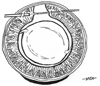
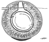
A 67-year-old woman presented to department of ophthalmology, Hanyang University Guri Hospital in April 2004 reporting visual deterioration in both eyes. Aware of her poor vision, the patient had no history of oculopathy, ophthalmic surgery, trauma, or disease. On initial examination, the manifest refraction was -1.50 diopters (D) with a best corrected visual acuity (BCVA) of 20/200 in the right eye and -2.00D with a BCVA of hand motion in the left eye. Intraocular pressure was 15 mmHg in the right eye and 14 mmHg in the left eye. A slit lamp examination with mydriasis revealed a corticonuclear cataract and dehiscence of the inferior zonules without phacodonesis or subluxation of the lenses. Fundoscopy showed a tessellated fundus with inferior chorioretinal coloboma that did not involve the macula or optic nerves in the right eye. We were not able to observe the retina in the left eye due to a dense brunescent cataract. Ascan biometry demonstrated an axial ocular length of 24.29 mm in the right eye and 25.38 mm in the left eye.
Under retrobulbar anesthesia, phacoemulsification was attempted on the left eye. Because the brunescent lens could not be phacoemulsified, we performed extracapsular cataract extraction (ECCE). During the operation, a posterior capsule rupture and a serious zonule defect was detected, so a scleral fixation of the IOL was performed.
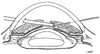
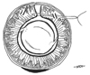
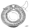
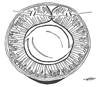
After two days, the procedures were done in the right eye by the same surgeon (J.H. Kim) according to a standardized protocol. Under retrobulbar anesthesia, a 3 mm sclerocorneal incision, paracentesis at the 2 o'clock position, CCC with a bent 25-gauge needle, and hydrodissection were performed uneventfully. Despite mild phacodonesis, the lens was normally located. Phacoemulsification was done in the bag using a stop and chop technique. Subsequently, high viscosity sodium hyaluronate was injected in order to expand the lens capsule completely. Because zonular weakness can cause lens decentration, a CTR was implanted through a sclerocorneal incision using a Sinskey hook. After the IOL (MA 60BM, Alcon®, USA) was placed centrally in the capsular bag, acetylcholine chloride 1:100 (Miochol®, Novartis®, USA) was injected through a side-port. Two paracenteses at 5 o'clock and 7 o'clock limbus were prepared for the iris repair. A long, straight needle (10-0 prolene Ethicon 1713; Johnson & Johnson®, USA) was introduced into the anterior chamber through the 7 o'clock paracentesis site. The right and left iris borders were picked up by the long, straight needle while the angled, blunt tipped 27 gauge needle, inserted from the 5 o'clock paracentesis, countered the pressure of the long, straight needle (Fig. 2) The straight needle was then inserted into the blunt tipped 27 gauge needle (Fig. 1). Both needles were pulled out through the 5 o'clock paracentesis (Fig. 3). After inserting the long needle into the blunt tipped needle from 7 o'clock, both were passed back through the 7 o'clock paracentesis site (Fig. 4). During this procedure, the blunt end of the long needle must be inserted first into the anterior chamber. If the sharp end of the needle gets into the anterior chamber first, it could go through the limbal tissue around the paracentesis and the thread could get caught on the limbal tissue. A single knot was loosely made (Fig. 5) and slowly tightened. The needles were pulled out again at the 5 o'clock paracentesis site to be tied (Fig. 6). Equal tension was used to tie both sides (Fig. 7). The knot was trimmed using vannas scissors through the sclerocorneal incision. This iris repair procedure was repeated three times.
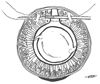
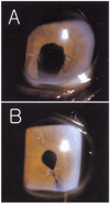
At 2 weeks postoperatively, visual acuity was 20/20 in the right eye and 20/40 in the left eye. In comparison with the left eye, the right inferonasal coloboma showed significant cosmetic and functional improvement (Fig. 8). At 3 months postoperatively, the IOL and pupil were centrally positioned in the left eye and no secondary problems were present.
Discussion
Two points should be considered regarding ocular coloboma during cataract surgery. Zonular and iris defects create potential misalignment problems of the pupillary center and the center of the intraocular lens.4 Moreover, iris defects may be associated with photophobia, and glare. We used CTR and repaired iris defects to improve this problem. The use of CTRs has been proven to be beneficial for IOL centration. When a zonular defect is present and a CTR is inserted at any stage of the procedure to reestablish the capsule's contour, the CTR protects against capsular fornix aspiration, consecutive zonular dialysis extension, irrigation fluid flow behind the capsule, vitreous herniation into the anterior chamber, IOL decentration, and closure of the capsular opening.5
We performed a phacoemulsification with CTR implantation in the right eye. The IOL was also centrally stabilized. In the case of Mizuno et al.6, before performing phacoemulsification, they inserted a CTR in order to perform the phacoemulsificaiton in a stabilized condition. However, in our case we inserted the CTR following the phacoemulsification. In contrast to Mizuno, we attempted to well centralize the IOL in the capsular bag.
Iris suture and prosthetic iris devices improve postoperative outcomes by reducing glare disability in iris deficiency and absence. An iris suture method is generally used, because prosthetics are generally implanted only in special situations and are very expensive.7 Therefore, we repaired the iris using the modified suture method.
Moreover, a small stab incision is made in the clear cornea at 6 o'clock to retrieve the thread at the point when most surgeons repair the inferior iris defect.4,8-10 In the modified McCannel suture method used by Blackmon, a long, straight needle was employed, similar to our method in limbus, but the thread was retrieved at 6 o'clock.10 In Siepser's slip knot method, tying was done on one side.11 However, these methods sometimes make it difficult to retrieve a part of the thread, especially when repairing a large iris defect. If the iris is excessively strained, iridodialysis may occur. Siepser's method requires a larger wound so that the thread can be pulled out. The surgical technique used on our patient differs from these techniques. We retrieved one end of the thread at the opposite site using a blunt 27 gauge needle, and tied it with same tension on both sides. Our technique has no need for an additional wound through which to pull out the thread because we use the cataract operation incision. Additionally, this method does not have a steep learning curve. This same technique can be used to repair other pupillary abnormalities such as those following trauma, previous sector iridectomies, and pupils that are permanently dilated.
We had to perform scleral fixation in the left eye because the posterior capsular rupture occurred due to a zonular defect during ECCE. Moreover, we did not restore the iris defect. Consequently, instability of the lens and a permanent iris defect remained, causing glare and a cosmetic problem in the left eye.
Despite this problem, at 3 months postoperatively, the IOL and pupil were well positioned in the center due to CTR and iris repair. The patient did not suffer from glare and has good visual acuity (20/20) in the right eye in comparison to the left eye (20/40). In conclusion, we recommend using CTR and iris suturing techniques for functional and cosmetic iris abnormalities in coloboma while performing cataract surgery.
 XML Download